Aorta

The aorta is a large, cane-shaped vessel that delivers oxygen-rich blood to your body. It starts in the lower-left part of the heart and passes through the chest and abdomen. Along the way, blood vessels branch off the aorta, extending to organs and supporting tissue.

Overview

Your aorta is the main vessel that takes oxygen-rich blood from your heart to the rest of your body.

What is the aorta?

The aorta is a cane-shaped artery. It starts in the lower-left chamber of your heart (ventricle). From there, it extends up toward your head a short distance before curving down. The aorta passes through your chest and abdominal cavities and ends at your pelvis.

Pairs of smaller blood vessels branch off from various points of the aorta. These branches extend the reach of the aorta to muscles, nerves and organs throughout your body.

Advertisement

Cleveland Clinic is a non-profit academic medical center. Advertising on our site helps support our mission. We do not endorse non-Cleveland Clinic products or services. Policy
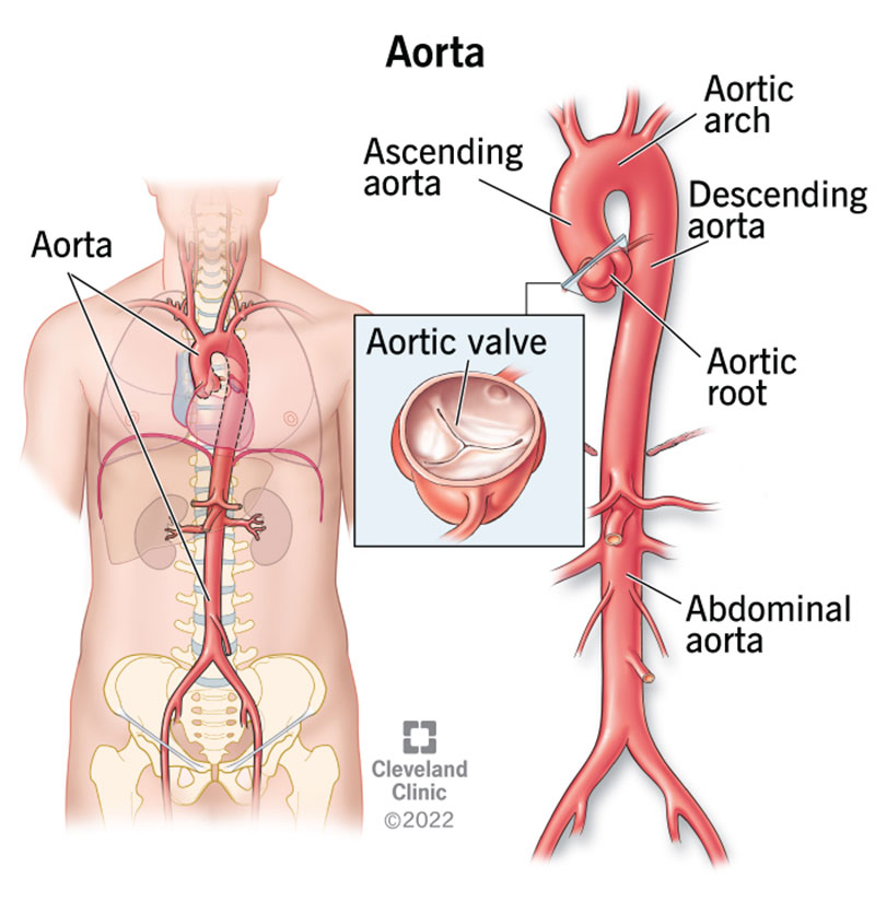
How large is the aorta?

The aorta is the largest blood vessel in your body. It’s more than 1 foot long and an inch in diameter at its widest point. As the aorta makes its way toward your pelvis, its diameter narrows to two centimeters.

What is the aorta made of?

It’s a complex structure that has three layers of tissue. These include:

  • Inner layer (tunica intima): This is the tube through which blood passes. It contains smooth muscle tissue, connective tissue and endothelial cells. These special cells enable blood to transport oxygen and nutrients without getting absorbed until it reaches the right spot.
  • Middle layer (tunica media): This layer is made of smooth muscle tissue, elastin and collagen (proteins). These substances enable the aorta to meet your body’s changing blood flow needs. When more blood is necessary, the aorta widens. If less blood is needed, it narrows.
  • Outer layer (tunica adventitia): The outer layer anchors the aorta in place. It also connects to nearby nerves and tissue.
Advertisement

Function

What is the function of the aorta?

The aorta is the main vessel through which oxygen-rich blood travels from the heart to the rest of the body. It also delivers nutrients and hormones. The aorta’s branches ensure these substances reach internal organs and nearby supporting tissue.

Why is the aorta’s function so important?

The aorta is the primary source of oxygen and essential nutrients for many organs. If disease or injury affects blood flow through this vessel, life-threatening complications can occur in minutes.

These include:

Advertisement

Anatomy

What is the anatomy of the aorta?

The aorta has many sections, including the:

  • Aortic root: Section that attaches to the heart. This is the widest part of the aorta.
  • Aortic valve: Three flaps of tissue (leaflets) that snap open and shut to release oxygen-rich blood from the heart.
  • Ascending aorta: Upward curve that occurs shortly after the aorta leaves the heart.
  • Aortic arch: Curved segment that gives the aorta its cane-like shape. It bridges the ascending and descending aorta.
  • Descending aorta: Long, straight segment that runs from your chest (thoracic aorta) to your abdominal area (abdominal aorta).

What are the aorta’s branches?

Many smaller blood vessels branch off from the aorta, including:

Ascending aortic branches

Aortic arch branches

  • Brachiocephalic trunk, which branches into the right subclavian artery (supplies the right arm) and right carotid artery (supplies the brain and right side of the head and neck).
  • Left subclavian artery supplies your left arm and the back of your brain.
  • Left carotid artery supplies your brain and the left side of the head and neck.

Descending thoracic aortic branches

  • Bronchial arteries supply the bronchioles, structures deep within the lungs.
  • Mediastinal arteries supply the mediastinum, a space between the lungs that houses the windpipe, esophagus and more.
  • Esophageal arteries supply the esophagus, a tube that connects the throat to the top of the stomach.
  • Pericardial arteries supply the pericardium, a protective sac that lubricates the heart.
  • Superior phrenic arteries supply the thoracic vertebrae, spinal cord and muscles, joints and skin of the middle back.

Descending abdominal aortic branches

  • Inferior phrenic arteries supply your diaphragm.
  • Celiac trunk arteries supply organs of your gastrointestinal system, including the stomach, liver, spleen and pancreas.
  • Superior and inferior mesenteric arteries supply your intestines.
  • Renal arteries supply your kidneys.
  • Gonadal arteries supply ovaries in women and testes in men.
  • Lumbar arteries supply the lumbar vertebrae, spinal cord, muscles, joints and skin of your lower back.
  • Median sacral arteries supply the lowest part of your spine (tailbone) and top of your pelvis.
  • Iliac arteries supply the lower extremities.

Conditions and Disorders

What conditions can affect my aorta?

Diseases, defects and injuries can affect the aorta’s ability to do its job. These conditions include:

Aortic valve disease

Diseases of the aortic valve include:

  • Aortic regurgitation (leaky heart valve): The aortic valve leaflets do not snap shut all the way. This causes blood to flow back into the heart instead of out to your body.
  • Aortic stenosis: The aortic valve becomes stiff and narrow, limiting blood flow out to your body.
  • Bicuspid aortic valve disease: The aortic valve has only two leaflets instead of three. This defect is present at birth but might not cause symptoms until adulthood.

Aneurysms, dissections and ruptures

Some of the more common issues affecting the aorta include:

  • Abdominal aortic aneurysm: A weak, bulging spot in the aorta that occurs as it passes through the abdominal region.
  • Dilated aortic root: Abnormal widening of the root of the aorta.
  • Thoracic aortic aneurysm: Bulge in the aorta that occurs as it passes through the chest.
  • Penetrating aortic ulcer: Fatty buildups (plaques) that break down the tissue of the aorta’s inner layer to reach the middle layer.
  • Aortic dissection: A tear in the inner lining of the aorta. This causes blood to seep into the space between the middle and inner layers.
  • Ruptured aorta: Tear that extends through all layers of aortic wall tissue. This causes oxygen-rich blood to spill out into the body.

Heart defects present at birth

Some congenital heart diseases affect the aorta, including:

  • Coarctation of the aorta: When the aorta is abnormally narrow. This makes the heart work harder than it should to pump blood out to the body.
  • Hypoplastic left heart syndrome (HLHS): A combination of heart defects affecting the left side of the heart. Children with HLHS have an aorta that’s too narrow to function.
  • Interrupted aortic arch: Rare condition in which the aorta doesn’t develop correctly. It affects blood flow out to the body and requires treatment shortly after birth.
  • Patent ductus arteriosus (PDA): Opening between the aorta and pulmonary artery that doesn’t close on its own after birth. This causes oxygen-rich blood to flow to the lungs.

Other types of aortic disease

Additional aortic conditions, some of which are rare, include:

  • Aortic atherosclerosis: Plaques that develop on the inside walls of the aorta.
  • Aortic infection: Rare condition that occurs when the aorta becomes infected.
  • Aortitis: Inflammation in the walls of the aorta. This rare condition is more likely to develop in people with autoimmune diseases that attack healthy tissue.
  • Aortic trauma: Severe injuries that tear or damage the aorta. These injuries may be due to falling from great heights, high-speed collisions or being ejected from a car.
  • Mural thrombus: Blood clots that affect blood flow through large vessels, such as the aorta.
  • Takayasu's arteritis: Inflammation affecting branches of the aorta that deliver blood to the arms, neck and brain.

Is aortic disease genetic?

Certain aortic diseases, including aneurysms and dissections, can run in families.

Genetic conditions, especially ones affecting the connective tissue, can also lead to aortic disease. These include:

Care

Am I at risk for aortic disease?

Your risk of aortic aneurysm, rupture or dissection increases with age. These issues are more likely to happen in older adults.

The risk of certain aortic diseases also increases when you have a chronic condition that damages blood vessels. These include:

Additional risk factors include:

  • Being male.
  • Drinking too much alcohol.
  • Smoking or using other tobacco products.

Does being at risk for aortic disease mean my health is in danger?

Not always. Conditions such as small aneurysms sometimes do not worsen. Others have symptoms that come on gradually.

If aortic disease symptoms are progressing, healthcare providers may recommend preventive therapies. For example, endovascular aneurysm repair is a non-surgical procedure that reinforces blood vessels to prevent rupture or dissection. This is one of many options that protect you against complications.

How can I prevent aortic disease?

Some conditions, including congenital defects, genetic diseases and trauma, are difficult to prevent. But there are steps you can take to avoid other types of aortic disease.

Prevention may include:

  • Adopting healthy living habits. These include eating a nutritious diet and being physically active.
  • Following care instructions for chronic conditions that damage blood vessels. Your healthcare provider may prescribe medications to lower high blood pressure and cholesterol or control blood sugar levels.
  • Limiting drinks containing alcohol to one or two per day or avoiding them altogether.
  • Managing stress.
  • Quitting smoking or other tobacco products.

Is there anything else I can do to avoid life-threatening complications?

If you are at risk for aortic disease or healthcare providers detect a minor issue, regular monitoring can help.

Services may include imaging studies and assessments from a vascular disease specialist. Sophisticated software, along with the specialist’s expertise, helps determine the likelihood of complications. The specialist can also let you know whether preventive treatment is necessary.

Imaging studies may include:

A note from Cleveland Clinic

Your aorta is a long blood vessel that’s essential to your well-being. It provides a steady supply of oxygen and nutrients to your organs. There are many conditions that can affect aorta functioning. Some can put you at risk for life-threatening complications. But having aortic disease or being at risk doesn’t always mean your health is in danger. Taking good care of yourself and seeing a specialist, when necessary, helps many people avoid complications.

Medically Reviewed

Last reviewed by a Cleveland Clinic medical professional on 02/24/2022.

Learn more about our editorial process.

Ad
Appointments 800.659.7822