The great vessels of the heart include your aorta, pulmonary trunk, pulmonary veins and vena cava (superior and inferior). They connect directly to your heart and play a vital role in your circulatory system. These blood vessels send blood between your heart and lungs (pulmonary circuit) and between your heart and body (systemic circuit).
Advertisement
Cleveland Clinic is a non-profit academic medical center. Advertising on our site helps support our mission. We do not endorse non-Cleveland Clinic products or services. Policy
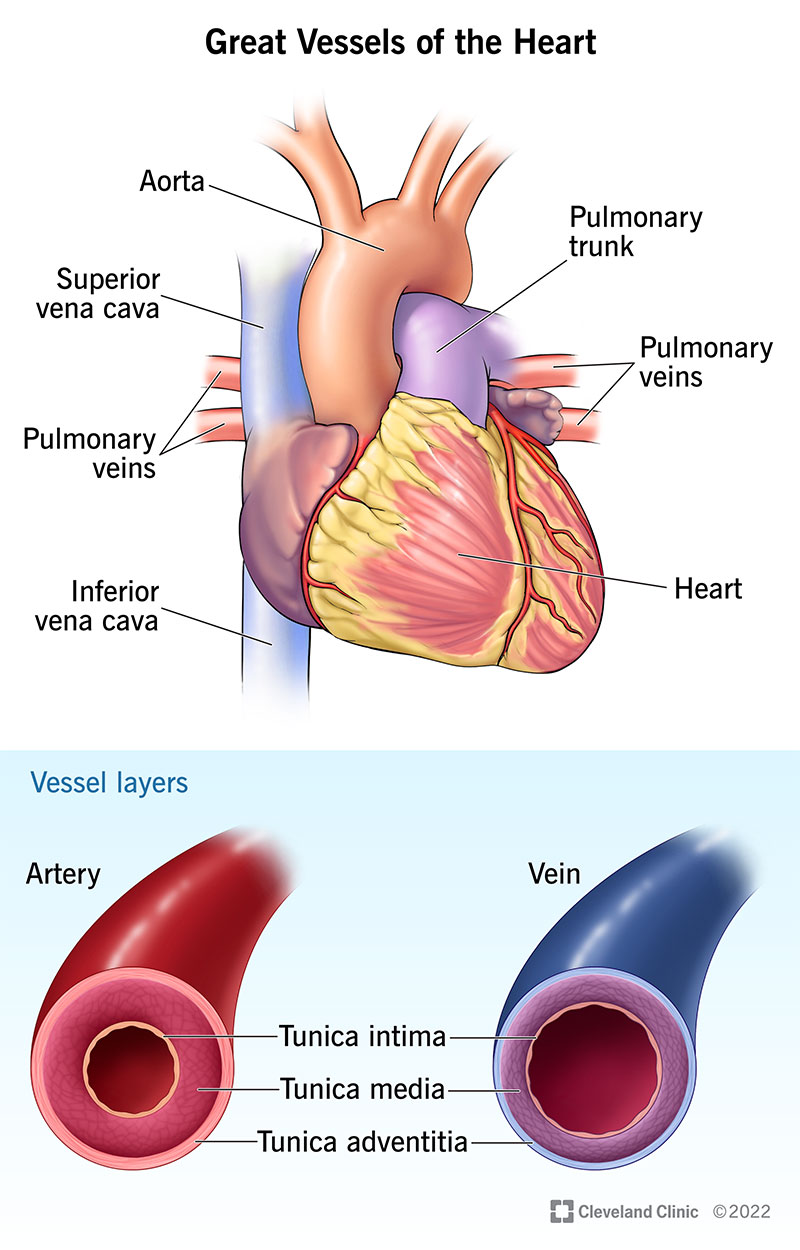
Image content: This image is available to view online.
View image online (https://my.clevelandclinic.org/-/scassets/images/org/health/articles/17057-great-vessels-of-the-heart.ashx)
The great vessels of the heart are major blood vessels that connect directly to your heart. These arteries and veins circulate blood between your heart and lungs, and between your heart and the rest of your body.
Advertisement
Cleveland Clinic is a non-profit academic medical center. Advertising on our site helps support our mission. We do not endorse non-Cleveland Clinic products or services. Policy
The great vessels include your:
Your great vessels work as a system of highways to keep blood moving in the correct paths throughout your body. These vessels connect with various chambers of your heart to send blood in and out of your heart in a coordinated fashion each time your heart beats.
Your great vessels are a vital part of your circulatory system. There are two main circulatory system circuits: the pulmonary circuit and the systemic circuit. Here’s a breakdown of what those circuits do and the role your great vessels play:
Your pulmonary circuit sends blood between your heart and lungs. First, oxygen-poor blood travels from your heart to your lungs. There, it receives oxygen and gets rid of waste. This refreshed blood then travels back to your heart.
The role of your great vessels
Advertisement
Your systemic circuit sends blood between your heart and the rest of your body. First, oxygen-rich blood leaves your heart. It circulates throughout your body, where it delivers oxygen, nutrients and hormones to your organs and tissues. It also picks up waste. This blood, now low on oxygen and containing waste products, travels back to your heart.
The role of your great vessels
Your great vessels are similar to other blood vessels in your body. The arteries carry blood away from your heart, and the veins carry blood toward your heart. However, there’s a crucial difference.
Normally, arteries contain oxygen-rich blood, and veins contain oxygen-poor blood. However, there are two exceptions to this rule: your pulmonary arteries carry oxygen-poor blood, and your pulmonary veins carry oxygen-rich blood.
The great vessels of the heart connect to your heart’s chambers. The chart below shows where each vessel connects and the direction of blood flow.
| Great vessel | Where it connects to your heart | Direction of blood flow |
|---|---|---|
| Aorta. | Left ventricle (via your aortic valve). | Heart to artery. |
| Main pulmonary artery. | Right ventricle (via your pulmonary valve). | Heart to artery. |
| Pulmonary veins. | Left atrium. | Vein to heart. |
| Superior vena cava. | Right atrium. | Vein to heart. |
| Inferior vena cava. | Right atrium. | Vein to heart. |
| Great vessel | ||
| Aorta. | ||
| Where it connects to your heart | ||
| Left ventricle (via your aortic valve). | ||
| Direction of blood flow | ||
| Heart to artery. | ||
| Main pulmonary artery. | ||
| Where it connects to your heart | ||
| Right ventricle (via your pulmonary valve). | ||
| Direction of blood flow | ||
| Heart to artery. | ||
| Pulmonary veins. | ||
| Where it connects to your heart | ||
| Left atrium. | ||
| Direction of blood flow | ||
| Vein to heart. | ||
| Superior vena cava. | ||
| Where it connects to your heart | ||
| Right atrium. | ||
| Direction of blood flow | ||
| Vein to heart. | ||
| Inferior vena cava. | ||
| Where it connects to your heart | ||
| Right atrium. | ||
| Direction of blood flow | ||
| Vein to heart. |
Most people have four pulmonary veins. They each drain blood from a different section of your lungs and carry it to your heart. They’re called:
Three layers of tissue make up the walls of your great vessels:
Like your other blood vessels, your great vessels have a tube-like shape. The walls surround and protect the lumen, or the opening through which your blood flows.
The great vessels of your heart have a wider lumen (opening) compared with your other arteries and veins. They need to be wider to accommodate the heavy volume of blood flow. Your aorta and pulmonary artery must also withstand forceful pressure from your heart’s pumping action.
Advertisement
The diameter (width of the lumen) varies based on many factors like your age and sex. Plus, different imaging methods establish different diameters in published research. The estimates below give you a general idea of the diameter of your great vessels:
Many conditions can affect your great vessels. These include congenital heart diseases (present at birth) as well as conditions you develop later in life. Listed below are the great vessels and some of the conditions that can affect each one:
Aorta
Pulmonary artery
Pulmonary veins
Advertisement
Superior and inferior vena cava
A heart-healthy lifestyle can help you keep your great vessels and all your blood vessels healthy. Tips include:
The great vessels of the heart are truly “great.” They play a major role in sending blood to and from your heart and supporting the daily work of your circulatory system. Your great vessels allow all your other blood vessels to do their jobs and supply your body with oxygen as well as remove waste.
Learning your great vessel anatomy can help you picture what’s going on inside your body with each heartbeat. Talk to your healthcare provider if you have questions or concerns about your blood vessels or what you can do to keep them healthy.
Advertisement


Sign up for our Health Essentials emails for expert guidance on nutrition, fitness, sleep, skin care and more.
Learn more about the Health Library and our editorial process.
Cleveland Clinic’s health articles are based on evidence-backed information and review by medical professionals to ensure accuracy, reliability and up-to-date clinical standards.
Cleveland Clinic’s health articles are based on evidence-backed information and review by medical professionals to ensure accuracy, reliability and up-to-date clinical standards.
When your heart needs some help, the cardiology experts at Cleveland Clinic are here for you. We diagnose and treat the full spectrum of cardiovascular diseases.
