Aortic Coarctation

Coarctation of the aorta is a congenital heart defect in which your baby’s aorta (the largest artery in their body) is pinched in or narrowed in one spot. This restricts normal blood flow to their lower body. Surgery or catheterization can repair the problem and relieve symptoms. Long-term follow-ups are essential even after repair.

Overview

What is coarctation of the aorta?

Coarctation of the aorta is a congenital (present at birth) heart defect. It’s also called aortic coarctation (pronounced “ay-or-tuhk” “koh-ark-TEY-shun”). This defect affects your baby’s aorta, which is the largest artery in their body. It carries oxygen-rich blood from your baby’s heart to the rest of their body.

If your baby has aortic coarctation, one part of their aorta is narrower than it should be. Picture a long balloon that’s used to make balloon animals for kids. You twist the balloon at one point to begin forming a shape. This causes the balloon to be pinched inward at that point. The pinch in the middle of the balloon is similar to what an aortic coarctation looks like.

That pinched point might be very narrow and cause severe symptoms soon after birth. Or it might be narrower than normal but wide enough to let blood pass through. In that case, symptoms might not appear until later in childhood or adolescence. Symptoms such as hypertension (high blood pressure) may lead to detection of aortic coarctation.

Babies who have coarctation of the aorta may also have other heart problems, including:

Aortic coarctation needs treatment to prevent serious complications. Once a diagnosis of aortic coarctation is made, either surgical repair or balloon angioplasty should be performed.

A group of illustrations showing the anatomy of the heart and aorta. Side-by-side pictures show a healthy aorta compared with one that's narrowed.

If your baby has aortic coarctation, that means one part of the aorta is narrower than normal. Usually, this narrowing occurs in the descending aorta.

How common is coarctation of the aorta?

About 4 in 10,000 babies in the U.S. have coarctation of the aorta. Each year, about 2,200 babies are born with the condition.

Of all babies born with some form of congenital heart disease, about 7 in 100 have aortic coarctation.

How does coarctation of the aorta affect my baby’s body?

Coarctation of the aorta prevents blood from flowing normally throughout your baby’s body. The narrowing causes the main pumping chamber of your baby’s heart (left ventricle) to pump harder than normal. This puts a lot of stress and strain on their left ventricle. In cases where the narrowing is severe, not enough blood will get to their body and their brain, which can cause shock and even death. In milder cases, this extra strain can cause your baby’s heart muscle to get thicker (hypertrophy).

Coarctation also causes higher blood pressure in your baby’s upper body, and lower blood pressure in their lower body. Your baby may have reduced blood flow to their kidneys, liver and other organs in their belly.

Advertisement

Cleveland Clinic is a non-profit academic medical center. Advertising on our site helps support our mission. We do not endorse non-Cleveland Clinic products or services. Policy
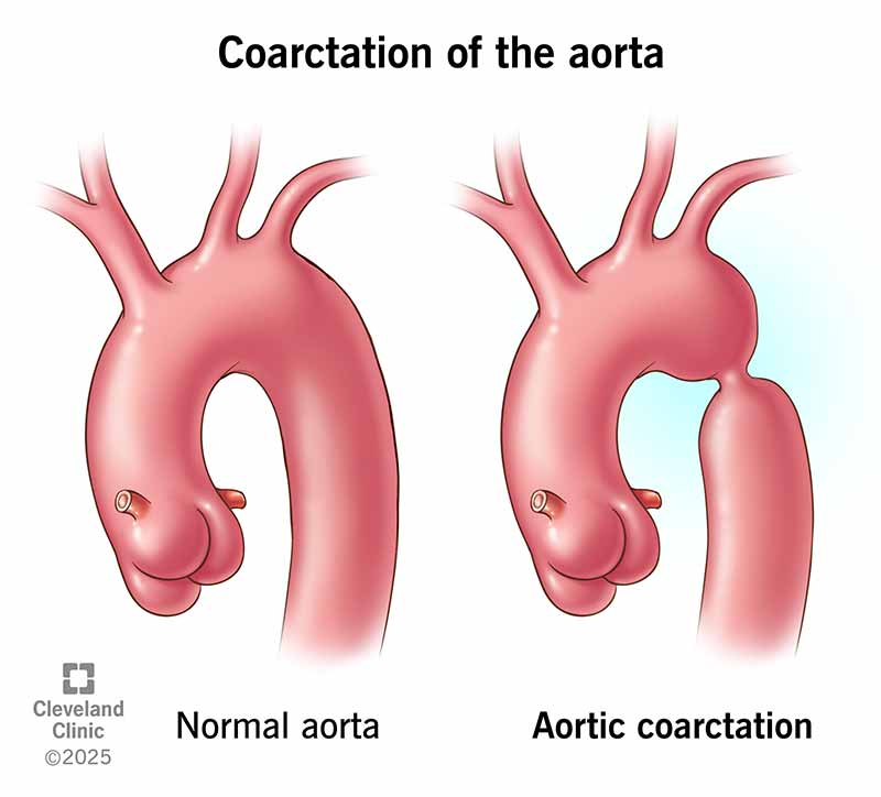
Symptoms and Causes

What are the symptoms of coarctation of the aorta?

The symptoms of aortic coarctation depend on how narrow the aorta has become. Some infants have no symptoms because the coarctation (narrowing) is very mild. But symptoms can appear within the first two weeks of birth if the coarctation is moderate or severe and the ductus arteriosus (a special fetal blood vessel) closes.

Symptoms of aortic coarctation in infants

Symptoms in infants include:

  • Fast pulse.
  • Fatigue.
  • Gray or pale skin.
  • Heavy sweating.
  • Irritability.
  • Rapid or labored breathing.
  • Trouble breathing.
  • Trouble with feeding.

Severe coarctation in infants can lead to shock and even death if not recognized and treated promptly.

Symptoms of aortic coarctation in children

Most children don’t have any symptoms. They’re usually diagnosed after their provider notices they have high blood pressure.

Some children may have symptoms if their blood pressure is too high in their upper body or too low in their lower body. These include:

Symptoms of aortic coarctation in adults

Symptoms in adults are usually due to recoarctation of the aorta. This is aortic narrowing that returns sometime after a previous repair. Symptoms may include:

  • Headaches.
  • Kidney problems.
  • Frequent miscarriages.
  • Lack of energy when using your legs.

But these symptoms are linked with many other health issues in adults. So, having these symptoms doesn’t mean you have aortic coarctation. Call a healthcare provider to discuss any symptoms you have. They’ll give you a physical exam and run tests to diagnose what’s wrong.

What causes coarctation of the aorta?

The exact cause of coarctation of the aorta isn’t fully known. However, congenital heart defects are often caused by genetic changes that happen before birth. Babies with Turner syndrome have a higher risk of aortic coarctation and other defects that affect the left side of their heart.

Sometimes, environmental factors play a role in causing heart defects, too. These are factors related to the birthing parent. They include:

  • Having diabetes.
  • Being infected with rubella while pregnant.
  • Using recreational drugs, especially cocaine.
  • Using certain prescription medications, including anti-seizure drugs.
  • Being over age 35.

The role of the ductus arteriosus

The ductus arteriosus may cause coarctation of the aorta. The ductus arteriosus is a small artery that connects the fetal aorta and pulmonary artery. It helps your fetus get enough oxygen-rich blood during gestation when their lungs aren’t working yet.

Once your baby is born, their lungs start working. That means they don’t need the ductus arteriosus anymore. So, it usually closes up within a few days of birth. But when it closes, something may go wrong. Some tissue from the ductus arteriosus may blend in with tissue from their aorta. When this tissue tightens to close up the ductus arteriosus, it may narrow their aorta as well and lead to coarctation.

Advertisement

What is the most common site of coarctation of the aorta?

Coarctation of the aorta usually occurs in your baby’s descending aorta near their ductus arteriosus. But it can also happen in other parts of their aorta. These include other areas of the aortic arch or further down in your baby’s chest or belly.

Your baby’s aorta is a long, curved blood vessel that arches upward from the top of their heart and then curves down through their chest and belly. It’s shaped like a walking cane with a round handle. The “handle” curves upward (ascending aorta) and then downward (descending aorta). The top of that curve is called the aortic arch.

The location of aortic coarctation matters because it affects your baby’s symptoms. You can think of your baby’s aorta like a main road that has lots of intersections. At each intersection, another artery or group of arteries connects with the aorta and carries blood in a different direction.

Aortic coarctation often occurs between two major intersections. The first is where arteries branch off to the upper body. The second is where arteries branch off to the lower body. So, blood leaves your baby’s heart and travels along just fine. It reaches the first intersection and easily flows into the arteries that lead to your baby’s upper body. Meanwhile, some blood keeps moving along in their aorta. And soon it runs into trouble.

It’s like when construction forces a five-lane highway down to one lane. Only so many cars can pass through. It's the same with your baby’s aorta. That pinched, narrowed part can only let a little blood pass through. So, it’s harder for blood to reach the second intersection, where arteries carry blood to your baby’s lower body. The coarctation slows down blood flow in those arteries.

This is why the blood pressure in your baby’s arms and upper body will be high. But the blood pressure in their lower body and legs will be low. This blood pressure difference is a red flag that leads to an aortic coarctation diagnosis.

Diagnosis and Tests

How is coarctation of the aorta diagnosed?

Coarctation of the aorta is usually diagnosed during infancy or early childhood. The timing depends on the severity of the symptoms. Infants with moderate or severe symptoms are typically diagnosed soon after birth. Infants with mild or no symptoms may not be diagnosed until later in childhood when they begin to have high blood pressure. Aortic coarctation is rarely diagnosed in adulthood.

Some newborns are diagnosed before they show visible symptoms. This happens when a pulse oximetry test shows low levels of oxygen in their blood. Low oxygen can be a sign of a serious heart defect like coarctation of the aorta. So, babies with low oxygen would then receive more tests to identify the specific problem.

Most babies and children are diagnosed with coarctation of the aorta when a physical exam reveals certain red flags. Signs that your baby or child may have coarctation of the aorta include:

  • High blood pressure in their arms and upper body, but low blood pressure in their legs and lower body.
  • Differences in their pulse when measured at their neck versus their groin.
  • A distinctive, harsh heart murmur that their provider hears when placing a stethoscope on their back.

Tests to diagnose coarctation of the aorta

When a physical exam raises red flags, your baby or child needs more tests to confirm the aortic coarctation diagnosis. These tests include:

Advertisement

Management and Treatment

How is coarctation of the aorta repaired?

Surgery is the gold standard for repairing coarctation of the aorta. But there are other treatment options, too. The best method for repairing aortic coarctation depends on many factors, including:

  • Your child’s overall health.
  • The precise location of the coarctation.
  • The size and severity of the coarctation.
  • Whether there’s an associated aneurysm or valve disease.

Babies and children diagnosed with coarctation of the aorta need the care of a congenital heart specialist. You’ll find such specialists at high-volume hospitals that treat congenital heart disease every day. If your baby was diagnosed with aortic coarctation, they should be evaluated by a cardiac surgeon at a major hospital. The surgeon will identify the best form of treatment and the best timing.

Babies with severe symptoms right after birth may need medication before having surgery. A medication called prostaglandin (PGE-1) is used to keep your baby’s ductus arteriosus open. This allows your baby to get enough oxygen and become stable enough for surgery. Some babies may need medication to help their heart pump.

Surgery to repair coarctation of the aorta

Surgeries to repair aortic coarctation can be divided into two major categories: those that can be done through an incision in your baby’s side (thoracotomy), and those that must be done through an incision in the front (sternotomy). The types of repairs done from a thoracotomy approach include:

  • Resection with end-to-end anastomosis. If the coarctation is relatively small, the surgeon can remove the narrowed part of your baby’s aorta and re-join the two ends. This is called resection (removal) with end-to-end anastomosis. It’s often the best surgical option to treat aortic coarctation.
  • Resection with extended end-to-end anastomosis. When there’s some narrowing of your baby’s aortic arch, the surgeon can remove the narrowed part of their aorta and join the lower part (descending aorta) to an elongated incision in their aortic arch. This is called resection (removal) with extended end-to-end anastomosis. It’s the best surgical option to treat aortic coarctation with associated transverse aortic arch hypoplasia.
  • Subclavian flap aortoplasty. This method involves making the narrowed portion of your baby’s aorta wider. The surgeon takes tissue from your baby’s subclavian artery (a nearby artery) and uses that tissue to widen their aorta. This repair is rarely done today because it sacrifices the artery that supplies blood to your baby’s left arm.
  • Patch aortoplasty. This method also widens your baby’s aorta. But the surgeon uses a patch made of synthetic material rather than tissue from your baby’s body. This isn’t typically done today because it carries a major risk of later dilation (aneurysm) of the patch.

Cardiac catheterization to repair coarctation of the aorta

Cardiac catheterization is a good option for older children who have mild coarctation. It’s also used for children and adults who have recoarctation (when the aorta narrows again sometime after repair). It’s less invasive than surgery. Options include:

  • Balloon angioplasty. The surgeon inserts a thin tube called a catheter into a vein in your child’s leg. This catheter has a balloon at its tip. The surgeon guides the catheter to the narrowed part of your child’s aorta and then inflates it. The balloon widens the aorta to allow blood to flow through more easily.
  • Balloon angioplasty with stent placement. This option involves balloon dilation (widening) of your child’s artery along with the placement of a metal mesh tube called a stent. The stent offers sturdy support to keep your child’s aorta open over time. This is better than angioplasty alone if your child has severe, long areas of coarctation. It’s also performed if your child had angioplasty but still has a blood pressure difference between their upper and lower body.

Complications of aortic coarctation repair

Complications of aortic coarctation repair include:

  • Recoarctation (recurrent coarctation). The same part of the aorta narrows again later on. The risk is 5% to 10% after surgery and 11% to 15% after either form of balloon angioplasty.
  • Aortic aneurysm. The walls of the aorta bulge outward.
  • Pseudoaneurysm. There’s a contained leak within the aortic wall.
  • Scar tissue. Scar tissue forms at the repair site and causes the aorta to narrow again. This happens most often in infants who had emergency surgery for severe coarctation.

Your child’s surgeon will explain the risks and complications in greater detail. It’s important to understand the possible complications. But it’s also important to know that aortic coarctation is essential and life-saving for your child. Without repair, aortic coarctation is fatal.

Can coarctation of the aorta be cured?

Coarctation of the aorta can’t be cured. Surgeries and interventions repair the problem, but there’s always a risk of recurrent coarctation. Even after treatment, people with aortic coarctation face a higher lifelong risk of cardiovascular problems.

Your child will need lifelong follow-ups with a congenital heart disease specialist. They’ll offer advice and keep an eye out for any possible issues.

Prevention

Can aortic coarctation be prevented?

There’s no specific way to prevent aortic coarctation. But prenatal care may impact your baby’s chances of having congenital heart disease. If you’re pregnant or planning a pregnancy, it’s important to:

  • Quit smoking and avoid drinking alcohol.
  • Avoid secondhand smoke.
  • Avoid using recreational drugs, especially cocaine.

Talk with your provider about any concerns you have. Genetic testing may be helpful if you or other biological family members have congenital heart disease.

Outlook / Prognosis

How long can you live with coarctation of the aorta?

Thanks to improvements in diagnosis and repair, people with coarctation of the aorta can live to at least age 60. In the past, the average life expectancy for people with coarctation of the aorta was just 35 years old.

Living With

How will coarctation of the aorta affect my child’s life?

Your child may or may not have restrictions on activities. If your child had aortic coarctation repair and there aren’t any persistent blood pressure problems, they may have no limitations. Check with your child’s provider to learn more about their specific situation and needs.

For six months following aortic coarctation repair, your child will need to take antibiotics before visiting the dentist or having some surgeries. This is to prevent an infection of the heart called endocarditis.

Additional Common Questions

How does coarctation of the aorta affect adults?

Coarctation of the aorta can put adults at a higher risk of cardiovascular problems. Untreated coarctation of the aorta is most dangerous. About 9 out of 10 adults with untreated aortic coarctation don’t survive to age 50.

But nowadays, it’s rare for someone to be diagnosed as an adult. Most adults with aortic coarctation had repair surgery or catheterization as a child. And they go on to live healthy and happy lives.

Still, adults with a history of aortic coarctation face a higher risk of:

If you had a repair procedure for aortic coarctation, you probably see your cardiologist regularly for check-ups. These are essential for making sure your heart is functioning well. You probably also think about your heart health more often than your friends who didn’t have surgery or catheterization when they were younger.

Life gets busy, though. And you may not always have the time or energy to cook heart-healthy meals or get in enough exercise. Plus, if you feel healthy overall, it’s easy to forget about what’s going on inside of your body that you can’t see. You shouldn’t let worry consume you, but it’s important to do whatever you can to reduce your risk for future problems.

Here are some steps you can take to keep your heart health a priority:

  • See your cardiologist every year for a check-up.
  • Take your medications as prescribed. This is especially important for managing your blood pressure.
  • Check with your cardiologist before having any surgery or procedure for issues unrelated to your heart.
  • Avoid strenuous activities like power weightlifting that cause your blood pressure to spike too quickly.
  • Add moderate exercise (like 30-minute walks) to your daily routine whenever possible. Talk with your cardiologist about the types of exercise that are best for you.
  • Take good care of your teeth and gums. The health of your mouth affects your heart health.
  • If you’re planning to get pregnant, talk with your provider about any associated risks. Many people with repaired aortic coarctation can have a healthy pregnancy. But pregnancy can be riskier if your aorta is still narrowed or part of your aorta is enlarged. High blood pressure also raises your risk of complications.

Thanks to medical advances, more babies with congenital heart defects survive into adulthood. That means an increasing number of adults are living with congenital heart disease. It also means there’s a growing amount of resources and support available to you. Talk with your healthcare team about how to connect with resources and meet other adults who are in your shoes.

A note from Cleveland Clinic

Each year, 1 in 2,500 babies in the U.S. are born with aortic coarctation. If your baby is that “1,” you might be wondering why this happened. Researchers can’t always know the exact cause of a congenital heart defect. What is known is that advances in medical care allow babies born with heart disease to live longer than ever before.

Early diagnosis and treatment are critical for managing aortic coarctation. Talk with your child’s provider about next steps for care and treatment.

Medically Reviewed

Last reviewed by a Cleveland Clinic medical professional on 08/28/2022.

Learn more about our editorial process.

Ad
Appointments 800.659.7822