Pectus Excavatum: Modified Ravitch Combined with Cardiac Surgery
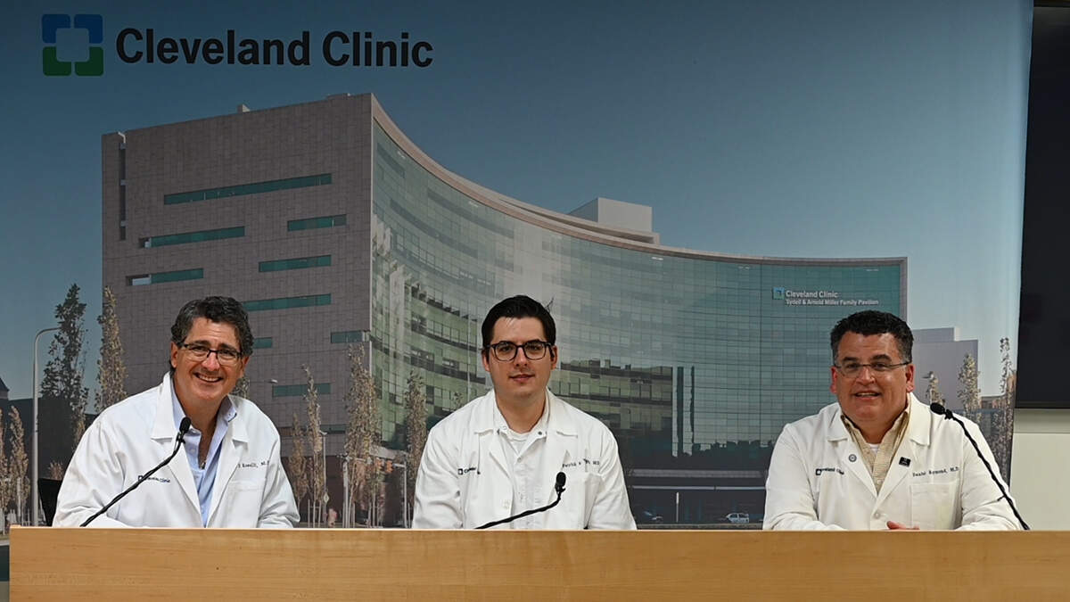
Some patients who have pectus excavatum also have cardiovascular problems, including congenital heart defects, valve disease, connective tissue disorders, and aorta disease. Repair of both problems in the same surgery can help patients feel better and look better with recovery from only one surgery. An experienced multidisciplinary team is necessary to determine the best treatment plan for the patient, plan the best surgery approach, insure the best recovery and have the best outcomes. Listen to Eric Roselli, MD, Daniel Raymond, MD, and Patrick Vargo, MD discuss their approach.
Reference: Anthony L. Zaki, Patrick R. Vargo, Dean P. Schraufnagel, Vidyasagar Kalahasti, Sudish Murthy, Eric E. Roselli, Daniel P. Raymond, Modified Ravitch Procedure for Pectus Excavatum Combined With Complex Cardiac Surgery, Seminars in Thoracic and Cardiovascular Surgery, 2021, https://www.sciencedirect.com/science/article/pii/S1043067921001088
Subscribe: Apple Podcasts | Spotify
Pectus Excavatum: Modified Ravitch Combined with Cardiac Surgery
Podcast Transcript
Announcer:
Welcome to Cleveland Clinic Cardiac Consult brought to you by the Sydell and Arnold Miller Family Heart, Vascular and Thoracic Institute at Cleveland Clinic.
Eric Roselli, MD:
Hello, everyone. I'm Eric Roselli from the Cleveland Clinic. I'm the surgical director of the Aorta Center and the chief of adult cardiac surgery, and I'm joined with my colleagues.
Patrick Vargo, MD:
My name is Patrick Vargo. I'm one of the heart and aorta surgeons here at Cleveland Clinic and a part of the Aorta Center.
Daniel Raymond, MD:
I'm Dan Raymond. I'm a thoracic surgeon at the Cleveland Clinic and head of the Center for Chest Wall Disease.
Eric Roselli, MD:
So it's great to be joined with, with these guys to discuss the topic of pectus excavatum. We together had written a paper recently looking at our experience in complex patients who present with the combined issues of a pectus excavatum, which is a chest wall abnormality that people are born with, where the sternum is depressed into the chest and cardiovascular problems. They often go hand in hand. What we've found is that by bringing our multi-disciplinary teams together, including other partners within the Heart, Vascular and Thoracic Institute, that we can provide comprehensive care to these patients. We wrote about it since we've had such a really great experience and a growing experience with it, and we're here to talk about these topics in more detail.
Eric Roselli, MD:
Patrick, Dr. Vargo, is a premier aortic surgeon here. You take care of a ton of patients with connective tissue disorder and complex aortic problems. Why don't you share with us a little bit of what you see when you're presented with patients who are referred to you with connective tissue disorders?
Patrick Vargo, MD:
Certainly. When we have a patient with a connective tissue disorder or somebody with a lot of the signs and symptoms of somebody with a connective tissue disorder, we'll bring them in for evaluation, both a comprehensive evaluation of their heart, aorta, major blood vessels, but also other aspects that go along with the connective tissue disorders. Marfan's, Loeys-Dietz, many of the others. We have a comprehensive center, like you said, and so they'll see folks from different areas that evaluate each of these, as well as genetic counseling, medical genetics, to see if we can identify what kind of syndrome they may have.
Patrick Vargo, MD:
Additionally, as part of their evaluation. Oftentimes we'll notice that they have a chest wall deformity as a part of the syndrome. If I find that they have a chest wall deformity while I'm evaluating them, I always pull in one of my colleagues like Dr. Raymond to take a look at the chest wall and give us his opinion about how it's affecting the patient. Is it significant? And if so, how can we address it in concert with their cardiovascular abnormalities?
Eric Roselli, MD:
And Dr. Raymond, Dan. See, we are all friends. I might use your first name here. Dr. Raymond, you have a special interest in the chest wall in so many ways from treating complex cancers to some of these congenital musculoskeletal abnormalities. Tell us a little bit about how this chest wall center kind of came to fruition, how your interest has grown around this topic.
Daniel Raymond, MD:
Well, I think it's pectus was one of the reasons why I really got interested in the chest wall deformity center and a chest wall center. We realized and actually interestingly in concert with the fact that we share clinic space with Dr. Roselli and Dr. Vargo, and we're always looking over each other's shoulders, looking at cases together. That was the genesis, the nidus of us starting to talk about, "Well, we're seeing a patient with pectus." "I'm seeing a patient with pectus, and I noticed they have an aortic abnormality." That created some synergy that built up the idea of, one, not only considering combined surgery, but also the broader issue of dealing with patients with chest wall disease.
Daniel Raymond, MD:
And the challenge there is that it's so poorly understood broadly even in medicine, and the classic pectus patient comes to us with a severe deformity, who's been told time and time again, "Oh, it's cosmetic, has nothing to do with physiology. You don't have a problem." And I have so many times seen people who are just completely incapacitated by this problem, and a simple surgery can really make a dramatic difference. And it's really exciting that then we can even extrapolate that to consider fixing two problems at once. So not having to put patients through two procedures, but one to address both problems at once.
Daniel Raymond, MD:
It was initially something that was approached with a lot of caution and trepidation, because you can create more problems. You can combine the surgeries and have bleeding issues. And that, as I think, what has limited the cardiothoracic community in general from exploring this, but we just have that unique synergy here where we realized we could do this. And we've finally been able to put together a series of patients in a paper that's showing that this can be done safely, and it can be done efficiently, and we can get very good results.
Eric Roselli, MD:
Yeah, it's one of the things. After being here at the Cleveland Clinic for 20, 24 years, I've been asked a lot of times, "Well, what's special about that place?" It really is the culture of collaboration. Not only are we all sort of in a center where our focus can be on the patient because of the construct business aspects and all these other things, but geographically in this Heart, Vascular and Thoracic Institute, it's just like you said, we operate next to each other, we see patients in clinic next to each other. And just like anything, you put two minds together, you get more than one plus one. That's been really fun. A lot of the patients with connective tissue disorder have musculoskeletal problems they have to deal with. Patrick, we even bring in people from other specialties to help with managing these patients, don't we?
Patrick Vargo, MD:
Right. Yeah. We do, and we consult our colleagues in orthopaedics and spine surgery as well oftentimes for an opinion and make sure they get fully evaluated for any kind of deformity that may not be in the chest, but that is affecting their lives.
Eric Roselli, MD:
Dr. Raymond, when someone has a pectus deformity, how are they affected by it physiologically? What are the symptoms that happen?
Daniel Raymond, MD:
I think many pectus patients will share with you the challenges primarily of limitations in their physiologic capacity. Essentially, there are two basic theories on why pectus is affecting people functionally. One is that with the sternum depressed, it's compressing the heart and preventing the heart from filling appropriately, and so there is limitation on the amount of blood that the heart can pump out with a single stroke. As a result of that, their reduced what we call stroke volume, their heart's functional capacity is reduced. And by alleviating that, by pushing the sternum forward, allowing the heart to expand more efficiently, we're allowing it to function more efficiently.
Daniel Raymond, MD:
The second thought is that there is a general loss of total chest volume that limits the ability of the lungs to expand. The way we see this is when people have pulmonary function tests, they have what's called a restrictive abnormality, meaning their lung volumes are smaller than expected. No, it's not. And the restriction is that the skeletal structure is small. Again, the pectus repair expands the intrathoracic volume, so that may be an additional means by which it benefits the patient physiologically.
Daniel Raymond, MD:
So how does that limit patients? They tend to have hit a plateau of exercise capacity that's lower than their peers. The person will go exercising or doing a treadmill, and the person next to them will be able to go 10 minutes, 20 minutes longer. I've had patients of the extreme who would exercise until they passed out, because that was they're pushing themselves to their limits of capacity. What do they feel when they're at their limits? Their heart is racing. They feel palpitations, and they simply just can't keep up with their peers. That is kind of a classic sign of pectus, and we can look at that more objectively by getting what's called a cardiopulmonary exercise test where we can actually quantify the amount of work that a patient can do based on the amount of oxygen their body consumes. What we find in the pectus patients is that they have abnormally low work capacity. So by fixing that pectus, we can increase that work capacity so that they can function similarly to their peers.
Eric Roselli, MD:
Do you do exercise testing on your pectus patients routinely as part of workup?
Daniel Raymond, MD:
I do routine exercise testing. That's one of the things we always tell patients when they come in, is to bring some exercise clothes so that they can go through the process that does involve getting blood gases. The one group that we don't necessarily do that in is patients who have underlying cardiac abnormalities that are being considered for cardiac surgery, because on the exercise test it can be very difficult to differentiate what is pectus limitation versus what is cardiac limitation. So there, we're basing it more on classic factors, such as the Haller index, which is the basic objective measurement that most people use.
Eric Roselli, MD:
What about postoperative exercise testing? Have you been able to demonstrate a benefit?
Daniel Raymond, MD:
We haven't done that formally. It's something where, honestly, I've been looking for the funding to do it as a postoperative test. We have in cases where people come back and don't think they've shown benefit, we've done exercise testing to show them that they've increased their functional capacity by 15 to 20%, which is about what you would expect, because that essentially oftentimes will normalize their functional capacity. It's a goal of mine in the future to have everyone tested pre- and post-op, it's just that I have to find the funding to pay for the tests postoperatively.
Eric Roselli, MD:
So this operation improves that sort of bucket handle, billowing mechanism of chest wall respiration.
Daniel Raymond, MD:
Exactly. Yeah. What's the downside of any surgery on the chest, is you lose some of what we call the compliance of the chest wall. The chest wall becomes a little stiffer. It's not free, but the surgery itself improves the intrathoracic volume, and then probably more likely, I suspect, it's the cardiac filling abnormalities, that we're preventing that right ventricle from being compressed. We're allowing the right ventricle to fill it to maximal capacity and allowing it to accommodate increasing needs as people exercise.
Eric Roselli, MD:
We definitely saw that in that first case we did together. It was remarkable. The woman couldn't walk to her mailbox. That was the story she told us, and she's just the sweetest thing. And then afterwards, after I fixed her tricuspid valve and closed her atrial septal defect and you reconstructed her chest wall, she was like running around with her family who are all these really active people living on a farm. That was like one of the coolest stories ever. We've got to give her credit for helping all these other people, because she was the one that pushed us to explore this.
Daniel Raymond, MD:
That was definitely kind of an epiphany moment when we saw her recovery and just how dramatically her life changed. I've had a few others. One is a woman who's been on YouTube who came to me with a fairly significant defect and was basically told all of her life that she was lazy and she just wasn't working hard enough. The change in her life after the surgery, it was life changing for her, and it was so gratifying to me. And it's really what has motivated me to push on this and really be dedicated to the practice population where a lot of people just don't take them seriously. It's something that, with experience, you can help people. You have to identify their physiological limitations and see if you can make them better, and it's very rewarding.
Eric Roselli, MD:
Yeah. It's not just a cosmetic benefit.
Daniel Raymond, MD:
No. No.
Eric Roselli, MD:
Although, you can't discount that. People are pretty excited about having sort of a normal looking chest well, for sure, afterward. I remember one guy was joking around how his brothers used to put cereal in-
Daniel Raymond, MD:
Yeah, the cereal bowl.
Patrick Vargo, MD:
The cereal bowl.
Eric Roselli, MD:
... the cereal bowl. Right.
Daniel Raymond, MD:
That's a common thing, yeah.
Eric Roselli, MD:
It's a funny story. But this operation is really interesting. The way that we conduct this is your team starts, clears out the cartilages, gives us exposure alongside the sternum, and then we work often in the left-side of the chest. Patrick, tell us a little bit about the orientation of organs in the chest of our patients, what it's like.
Patrick Vargo, MD:
Right. Right. Ordinarily, the heart sits right behind the sternum, and it's also towards the left of the heart. With a severe pectus or a severe deformation in the chest wall, it's often pushed to one side, and many times that is towards the left-side of the chest. So when we do these surgeries together, we said we first expose to the side of the sternum and remove the cartilage that's deformed. We'll actually shift the whole sternum sideways and have really excellent access to the heart structures. And within the series of patients that we've done these together in, we fixed the majority have had heart aortic operations, aortic repairs in the root, ascending, and even the arch. And the next most common operation was the mitral valve, which even in a normal patient can be ordinarily difficult to view sometimes, and there was excellent exposure with this approach. So going next to the sternum and moving the sternum over provided an excellent view of the cardiac and aortic structures below the chest wall that we needed to see to fix.
Daniel Raymond, MD:
What I would add to that is we've done the breadth of operations. We have a relatively small series, but we've still been able to do everything from a tricuspid valve repair, an ASD repair, a triple valve, a total arch, a Ross procedure, a David procedures. So this exposure is not limiting, but you have to have courageous cardiac surgeons who are willing to do things a little differently the first time. We had to tweak the technique, and there are ways of adapting the technique to provide different exposure at different levels. One of the things that's kind of unusual afterwards is the classic cardiac surgery patient has sternal wires. After this combined surgery, the patient doesn't have any sternal wires. So it's often people will look at a chest x-ray and say, "Ah, you didn't have cardiac surgery." It makes things a little different for the patients, and I always warn them now. I'm like, "People aren't going to believe you had cardiac surgery, just because they don't see that telltale sternal wire on your chest x-ray.
Eric Roselli, MD:
Yeah. There is a midline scar, though. Right?
Daniel Raymond, MD:
Yep, there is. There sure is.
Eric Roselli, MD:
The skin incision is still in the middle. One of the things, it's not just all of us coming together and talking about these things, but it's also what's made this available to us, I think, is the imaging technology, right?
Eric Roselli, MD:
We can take a CAT scan and do basically a virtual view of what the various sort of approaches might provide us. We can sit down with the 3D workstation and really get a sense of exactly how that incision needs to be, what portion of the chest wall needs to be addressed, what the structures are and how the structures are going to be exposed underneath. That's been a really nice tool for us to use.
Daniel Raymond, MD:
And that's been reflected in the fact that we don't have one single approach to this. There's a T-incision where there is an upper sternotomy, or there is a completely left parasternal approach. That is a team decision that's made preoperatively based on what is going to be the best exposure.
Eric Roselli, MD:
Yeah. And there is some variability, just like there's variability in everybody and everything.
Daniel Raymond, MD:
Absolutely.
Eric Roselli, MD:
I've been particularly paying a lot more attention when I see patients who have pectus, because there's a lot of patients have a mild pectus that we leave alone.
Daniel Raymond, MD:
Yes. Yep. Right.
Patrick Vargo, MD:
Mm-hmm (affirmative). Yeah. Right.
Eric Roselli, MD:
We'll leave it alone and do our cardiac operation, but I look a lot closer on it now and see that there is quite a bit of variability in where that ... I guess it's like a hinge point where you see the depression.
Daniel Raymond, MD:
Yep. Absolutely.
Eric Roselli, MD:
It's interesting that patients with connective tissue disorders like Marfan's, they often have a long chest wall, and they had sort of variable insertions of their ribs
Daniel Raymond, MD:
Right.
Eric Roselli, MD
Dan, does that change the way you think about how you're going to-
Daniel Raymond, MD:
No, I think one of our initial concerns with the connective tissue disorder patients was we do the typical ... The left parasternal approach is the Ravitch procedure, which most people will recognize the names. One of the concerns was, would they have any problems with the cartilage regrowing after we've done the left parasternal approach and divided the soft tissue, and then sewn it back together?
Daniel Raymond, MD:
That was one of the reasons why we truly waited on doing this paper until we had good, reasonable long-term follow-up with reasonable imaging, because all of the Marfan's patients get serial CT scans after surgery so we can look at their cartilage regrowth. We noted that they all regrew. There weren't patients that had defects in their chest wall and a billowing or a flail chest. I think in the connective tissue disorder population, that was my biggest concern that has been shown to be not a concern. Although the chest is longer, the sternum can be more blunted, and then the cartilage comes in in a more crowded, fused, abnormal fashion, which makes harvesting the cartilage and preserving the lining in the perichondrium a little more challenging. One of the things that I'm very pleased with this study that we've been able to do is look at the regrowth of the chest wall afterwards and show that this procedure didn't impact that.
Eric Roselli, MD:
Yeah. Yeah, I was a little worried about that last element you were talking about, that it might affect diaphragm function or something, but that really hasn't been an issue.
Daniel Raymond, MD:
No.
Eric Roselli, MD:
Two of the big concerns that people have about this idea of combining the things are, number one, whether there's an issue with the potential for more bleeding, and number two, pain management. Dr. Vargo, can you speak to the bleeding issue
Patrick Vargo, MD:
Yeah. We looked through it, all the blood transfusions and blood products that patients received. There were patients who did receive transfusions, but when looking at what they received, it wasn't out of the realm of what we see with complex cardiac and aortic surgery. We didn't have a direct comparison group to compare them. But looking at it, that we take care of patients that we do these operations in with and without a pectus, and the level of transfusion seemed reasonable.
Eric Roselli, MD:
Yeah. I thought that was true too, even with circulatory arrest.
Patrick Vargo, MD:
Right. And another important thing was, is that we didn't see any postoperative bleeding. A concern would be that with a more extensive chest surgery, that they would be at risk for bleeding after surgery, and that didn't seem to be the case. Patients did well after surgery. There were no re-explorations or any kind of bleeding events. It was a reasonable postoperative course.
Eric Roselli, MD:
Yeah. That's certainly relevant, because when you have to take somebody back in an emergency situation or consider doing CPR or something like that afterward, that can sort of give the team a little pause. I don't know if it's just us being more meticulous, or maybe our exposure is just really good through that broad exposure. Dan, can you comment about the pain?
Daniel Raymond, MD:
Yeah, pain control is a huge component of it. It's one of the biggest things when I talk to patients about recovery from a pectus surgery. What's unique about the combined approach is with a pectus alone repair, we like to use an epidural for temporary pain management. We can't place an epidural the day of surgery because of the anticoagulation required for the cardiopulmonary bypass. What we can do is, one, do regional blocks, and two, we can have an epidural placed once the coagulation parameters are normalized after surgery. So it's something where many of the patients get an epidural the morning of post-op day one. So the pain management again, which the bleeding and the pain management were our two major concerns, we've been able to work out solutions to all of those problems in a very satisfactory manner. And again, I think that works on our team approach.
Daniel Raymond, MD:
What I would say about the bleeding is certainly that was, again, one of our areas of trepidation in going into this, and I think that's one of the obstacles that a lot of people think about. That just took caution, time, and then it's observation afterwards. We're finding that we're not having excessive bleeding issues after the surgery.
Eric Roselli, MD:
Yeah. I've been impressed with the pain management. I guess, in general, we've been working on enhanced recovery after surgery pathways to help optimize our pain management. It looks a little different for cardiac than thoracic, but we've used all of those tools from both of our experiences and have been really impressed. Gosh, we had one patient who was out of here in like four or five days. She was laughing at the ... We were all worried about it. We warned her about pain, and she was laughing at us afterwards. She was like, "This is nothing, you guys." I don't know. Maybe she was comparing it to childbirth or something.
Daniel Raymond, MD:
The last one we did actually, when I went to see the patient on day three or four, his biggest complaint was the epidural hurt, which I've never heard anyone say that the epidural bothered them. For him to say that a little IV in the back hurt was very telling to me that the pain control was excellent, because he had no complaints about it. He could cough vigorously. I was very excited, because, to me, that's the patient who's going to do great.
Eric Roselli, MD:
Right.
Daniel Raymond, MD:
We can minimize their narcotic consumption. We can get them through the recovery faster.
Eric Roselli, MD:
Yeah. Well, this has been a really great discussion about one of the specialized things that we do here really well, and so I've enjoyed talking with you guys. Can you maybe give us some closing comments to our patients who are facing these problems and the caregivers taking care of them? Can you each give us some closing comment, Patrick and Dan?
Patrick Vargo, MD:
I think what's important is that many of the patients that come with these chest wall deformities have multiple issues going on that are part of a bigger syndrome of connective tissue disorders. And having a center here with many different specialists in different areas of the body from chest wall, aortic, ophthalmology specialists, spine specialists, we really can provide a comprehensive approach to that. I think our experience with fixing these defects, the pectus defect in combination with cardiac surgery has been shown to be very effective and safe to do. I think it's a valuable tool to offer patients to make them feel better and to have more energy and exercise capacity to enjoy their lives.
Daniel Raymond, MD:
A message to the broader pectus population. We hear you, we believe you, and we want to help you. I would strongly encourage people who have questions, who want evaluation, you should be seen at an experienced multidisciplinary center that has significant number of evaluations under their belt and can manage these complex issues. It is not just a cosmetic problem, and there are solutions for you. We really encourage you to pursue that. Get opinions, one opinion, two opinion, five opinions. Find a physician who listens to you, and that's one of the things I think we do very well here as a team, and we can provide excellent multidisciplinary care.
Eic Roselli, MD
Fantastic. Thanks a lot, guys.
Daniel Raymond, MD:
Thank you.
Patrick Vargo, MD:
Thank you.
Eric Roselli, MD:
As always, enjoyed working with you.
Announcer:
Thank you for listening. We hope you enjoyed the podcast. We welcome your comments and feedback. Please contact us at heart@ccf.org. Like what you heard? Subscribe wherever you get your podcasts or listen at clevelandclinic.org/cardiacconsultpodcast.
Cardiac Consult
A Cleveland Clinic podcast exploring heart, vascular and thoracic topics of interest to healthcare providers: medical and surgical treatments, diagnostic testing, medical conditions, and research, technology and practice issues.


