Redefining Care: Endoscopic Skull Base Surgery and Beyond

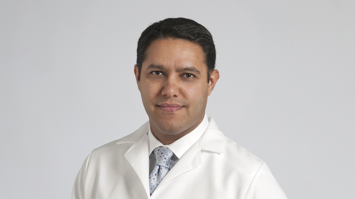
It's our 50th episode of Head and Neck Innovations! Returning guest Raj Sindwani, MD highlights the practice-changing approaches to complex endoscopic skull base surgery and post-operative monitoring that are part of Cleveland Clinic's plan of care.
Subscribe: Apple Podcasts | Spotify | Buzzsprout
Redefining Care: Endoscopic Skull Base Surgery and Beyond
Podcast Transcript
Paul Bryson: Welcome to Head and Neck Innovations, a Cleveland Clinic podcast for medical professionals exploring the latest innovations, discoveries, and surgical advances in otolaryngology-head and neck surgery.
Thanks for joining us for another episode of Head and Neck Innovations. I'm your host, Paul Bryson, Director of the Cleveland Clinic Voice Center. You can follow me on X, formerly Twitter, @PaulCBryson, and you can get the latest updates from Cleveland Clinic Otolaryngology-Head and Neck Surgery by following @CleClinicHNI on X. That's @CleClinicHNI. You can also find us on LinkedIn at Cleveland Clinic Otolaryngology - Head and Neck Surgery, and Instagram at Cleveland Clinic Otolaryngology.
Today I'm joined by a returning guest, Dr. Raj Sindwani, a rhinologist and Vice Chair of Head and Neck Operations for our Department of Otolaryngology-Head and Neck Surgery. He has been an innovator and leader of our endoscopic skull base surgery program for many years, and I’m just pleased to welcome you back to the podcast.
Raj Sindwani: Thanks for inviting me back, Paul. I'm glad to be here.
Paul Bryson: Well, for our listeners who maybe have not had the chance to go back and listen to our prior episode, please start by just sharing a little bit of your background, where you're from, where you trained, how you came to Cleveland Clinic, and maybe just set the table for the evolution of the endoscopic skull base program as you've been here.
Raj Sindwani: Sure. So first, those listeners should go back and listen to those other podcasts. Agreed. Agreed. You've had some great guests and a lot of interesting discussions and I've enjoyed listening to them myself.
So as you know Paul, I came to the Cleveland Clinic back in 2010 having done my med school and residency in Canada and fellowship in Boston at the Massachusetts Eye and Ear Infirmary many moons ago. I was in St. Louis for a couple of years and then got recruited here to kind of build out the rhinology section, with a big part being changing the landscape of endoscopic skull base surgery. It wasn't until I partnered after being here for a year or so with Dr. Pablo Recinos, the other co-director for the endoscopic skull base and minimally invasive cranial base program that we have here. And it's amazing to think back because prior to 2011, circa 2011-2012, even at the world famous Cleveland Clinic, as in many, many institutions, routine pituitary surgery was being done yes, trans-nasally, but with a microscope and an old school hearty speculum. Once I met my better half Dr. Recinos, we really wanted to change that paradigm, and serendipity played a big role. Several old school surgeons left the Clinic, and it left us with a huge runway to capitalize on some of the more minimally invasive techniques. So now as you know, we take out not just pituitary tumors, but a lot of aggressive and varied intracranial pathology going through the nose, using it as a tunnel or a conduit to get to pretty interesting places within the head.
Paul Bryson: Yeah, I mean, it's been amazing to see the growth that you guys have been able to achieve, and I'm always, we're going to talk a little bit about it, but I'm just really impressed with how the corridors have changed. It started with pituitary adenomas, but that's just sort of table stakes now for endoscopic skull base surgery. So there's, we're going to talk a little bit about it, but that's just like one door…
Raj Sindwani: It's one door and one avenue, and you're, so right now we actually use what's called multiport surgery going through the cheek sinus, for example. And we actually have kind of transposed some of that skillset and learning that we did by going through the nose to get to the intracranial cavity now to also access other no man's lands like the orbit, for example. So whether it's a trans-orbital technique to use the orbit itself as a corridor, or using the orbit as a target because there's pathology there, a tumor, whatever the case may be, we absolutely have been using that same two surgeon, multi-hand, multi-port approaches to get to a lot of different areas. So thanks for bringing that up. You're exactly right.
Paul Bryson: Well, I know earlier this year you and the team were at the North American Skull Base Society's annual meeting, a constant presence there over the years I've observed. Can you share a little bit of some of the things that you're excited about? And just as a consumer of your research and the team's research, I was specifically interested in some of the work that you're looking at for unplanned reoperation in skull-base surgery, and even tell me a little bit about idiopathic intracranial hypertension, something that when I was in medical school, really I wasn't aware of, was managed surgically very much, certainly not through the nose.
Raj Sindwani: Absolutely, yes. We can touch on those too. That's too of a whole host of research projects that myself, the team, our army of great med students and fellows presented, and that NASBS meeting is special to us because that's actually the only meeting of the year where both the Neuro and the ENT or Rhinology sides come together. So it's actually, we roll pretty deep at that meeting because all the fellows from years past on both sides come out to meet friends and to connect with the family.
So you mentioned the unplanned re-operation after endoscopic skull base surgery. This hasn't been looked at much in the literature, at least to the extent of what happens after the first few weeks after having endoscopic skull base surgery. How often do people have to go back to the OR? And our specific question was, why do they go back to the OR within, let's say that first year out after tumor removal specifically?
So we looked back at our five-year experience of over 500 patients, and what we found is that 5% of those patients actually did have some sort of endonasal surgery within that first year after surgery. Most had it within 30 days, and the median was two weeks. And when we dug deeply into what was actually taking people back to the operating room, not surprisingly 50% or so was because of postoperative CSF leak. That usually happens early after the surgery. As we all know, my interest was actually on what the other categories were, and surprisingly about the next most common reason to have someone go back to the operating room was actually residual or recurrent mass within that first year for a variety of pathologies. And then the next after that was hematoma or hemorrhage. Hematoma and hemorrhage also tended to happen early on after the procedure, and then although a small number of about 5%, but a big reason to us was patients were going back to the operating room that first year after endoscopic skull base surgery for sinus related issues, early mucus EAL formation obstruction of one sinus or the other, which we thought would be related to the approach.
Now we have evolved in our approach to the skull base, both routine pituitary surgery and really anything that's within this sphenoid proper. Unlike many centers, we do not do formal ethmoidectomy on both sides, and we do not routinely take the middle turbinate either. There's a few reasons for that. One, we make very small openings in the back of the nose so that we can access through this foid interior and the middle turbinate really we feel now that we've evolved in our facility with these techniques isn't necessary to be removed, meaning we can get the same exposure if the middle terminate is there or not. So our reason for keeping it is one, it's obviously a normal structure has a physiologic role, it has a neuro epithelium for smell, etc.. But one other reason is that it actually can serve as the guardian to a skull-based defect.
There actually is a case report of a patient dying because an NG tube was placed through the nose, unbeknownst into a skull-based defect. In this case, the patient became paraplegic and died. So in other words, when you expose all of these amazing intracranial structures and just put some mucosa over top of them, that's still a very delicate area of the body. So what we do is make a small hole, achieve still the great results through the nasal approach, but then keep the middle and actually put them against the septum. We did a cadaveric study a few years ago that actually showed when you do that blinded insertion of Dobhoff or NG tubes are greatly limited as far as whether they can get into this or not.
Paul Bryson: It sort of creates the channel for where the tube should go. Most of the time they're passed blindly.
Raj Sindwani: Exactly. Bounces off this middle turbinate face, goes right to the nasopharynx when you want it. And we had just totally random people, a med student, a junior resident, whoever we could find to serve as these blind tube passers in that study, which was kind of fun and it showed what we thought it would. It's important to keep that middle tube if you can.
Paul Bryson: No, that's a great one. That’s a fun one.
Raj Sindwani: You also asked about another project that we are really proud of, and this really builds on a thread of sort of some work we've been doing for many years. A few years ago, we became really fascinated with our results on lateral sphenoid sinus meningoencephalocele management. So traditionally these were managed through what's called the transpterygoid approach you go through the back wall, the maxillary sinus, you dissect through that complex pterygopalatine fossa contents and land up right directly into the lateral recess. Well, that PPF or pterygopalatine fossa has some very important structures, right? Nerves, blood vessels, arteries, et cetera. And the thought was why do you have to go through all that important anatomy when we're so facile using endoscopes that are angled and have all these specialized angled instruments? So we published one of the largest series of lateral encephalocele cases where they were managed through what we coined as the modified transpterygoid approach. So in a nutshell, we keep the PPF contents intact, remove the bone in front of it and behind it and mobilize it such that we can now use an angled scope, 30 or 45, look around the corner and patch the lateral sphenoid and encephalocele. Amazing success rates with that, I don't think we had even one failure in that series, and we were able to preserve all of that anatomy and importantly, minimize the morbidity of things like V2 numbness, bad bleeding, dry eye, and things like that.
Paul Bryson: That's amazing. I mean, in the contents of things that you would just traverse acts almost like a little patch on top of the patch for you when you…
Raj Sindwani: That's right. Yeah, it can if you end up filling with fat or something. So in that series, the large majority were patched only with a free graft too. So we were able to really deescalate how complex the approach was and therefore the reconstruction as well. So regardless of how you approach these lateral or even cephaloceles that you might get through the middle cranial fossa in the ear, we all know that when they occur spontaneously in women in their forties who may be obese and fertile, we all remember all those clinical kind of important salient features of that you still have to look for and manage potentially IH idiopathic intracranial hypertension, formerly known as benign intracranial hypertension. So following along that line of inquiry, okay, we're great at now patching these leaks, et cetera, but why are people getting these leaks to begin with and how concurrently do we manage the IIH in these patients?
And so this NASBS pass, we presented our management algorithm for IH specifically in skull-based meningoencephalic seals. It was a large group of patients, 80 patients or so, 50 of whom had really great follow-up of intraoperative opening pressure measurement, intrahospital measurements. So a few days later prior to removing the lumbar drain and then a six week opening pressure measurement also, we wanted to learn what the dynamics of the CSF pressure was. And what we found was pretty interesting. One was that using our algorithm, what we ended up kind of doing is that if people had an opening pressure of less than 20, they were not treated with anything, just counseled on weight loss. If they had an intermediate opening pressure, 20 to 30 millimeters of water, they're treated with acetazolamide or oral lowering agents for ICP. And if it was higher than 30, that's when we had the discussion of shunting them.
So 20 and 30 being the key yard post for when we would escalate our treatment. What we found is that when you looked at these 50 patients where we had all of these measurements, interestingly, when you put a patch in a patient's who's leaking post-op, it doesn't mean that their pressure will necessarily go up. Old surgical dogma was, well, you have an open system, so their pressure's low. If I put my finger in the dike, now that pressure's got to go somewhere, is that pressure going to be elevated? And our study suggests, no, it's not actually elevated. And furthermore, when you go out to that six week poke or opening pressure measurement, 23% of patients actually ended up going into the higher risk group than what they were in, and 35% or so went to a lower risk group than they were originally stratified to important, because as I just mentioned, if you get to a high risk group, you may need VP shunting. So this was some surprising data that we found. One, we did have an excellent success rate using this algorithm. We only had a few people that did leak, some early, some late, and we think the IH may be a very central component in some of those late failures. But what it also showed us is that we do need to continue to learn and get more data on how CSF dynamics are at play in this very special patient population.
Paul Bryson: Yeah, it's really interesting. Are the patients pretty varied or is it kind of like you led with, there's a mnemonic for who are these patients? When you look at that 80 patient group, are they from all backgrounds or do they kind of fit the mold that the old books tell us?
Raj Sindwani: Yeah, they do fit the mold and certainly we know clinically our antenna go up when you see that female in her forties, et cetera. But there were some men in there as well that still had IH. So it's not that it's exclusively that group, but it is something that you still need to keep your eyes on. The big question for us is, okay, now that we've identified it through the different stigmata that we see on MRI and so on, what do we do about it? It's interesting. I was part of one of the authors on a position, international consensus statement on the management of CSF leak spontaneously, and the group decided to actually veer around this topic of IH measurement and management because we couldn't come to a consensus on that topic on when do you measure opening pressure? Do you measure opening pressure because of a host of reasons including that we need to have more information on it?
Paul Bryson: Well, it sounds like that's going to evolve. You're going to keep looking for sort of signal refinement there and refining the algorithm.
Raj Sindwani: Exactly.
Paul Bryson: Well, I wanted to also, you'd kind of talked about all the different windows and portals and reconstructing them and things like that. I also wanted to give a little time for you to talk about the evolution of maybe some fascial reconstruction. You mentioned the workhorse nasal septal flaps and things, but if I recall, I understand that fascia lata is also a reconstructive ladder option for you in some of these complex skull-based defects. Can you give us an update on that?
Raj Sindwani: Sure, absolutely. You're right. So while the nasal septal flap is the workhorse for pedicled reconstruction, and we use that for large complex tumor defects that often have high flow leaks to them, that inner layer, what you do as the first layer in these multi-layer reconstructions has been shown to be very important. So we really have turned to fascia lata in our expanded, more complex, think of large defects, complex aggressive pathologies like cranial pharyngeal, sometime meningioma or other tumors. And what we've found, and this was a large series that we presented a little while ago that's now been published in 2023, is that when we looked at this large series of consecutive expanded approach surgeries for the reasons I mentioned, when we use fascia, we had a higher than 92% success rate. And what we've learned from that is that the fascia really is something special.
Compare it to the omentum in the abdomen. There's something about it in addition to just its physical characteristics, we harvest fascia from the upper thigh through a small incision, and it's the iliotibial band that we're effectively taking. And so we've been using it in a variety of different forms, which has also been a great learning experience. The button graft was popularized some years ago. The idea here, Paul, is if you think about it, you make a big hole, and if there's a big tumor cavity that's now just open space, how do you make your inner layer stay at the level of the skull-based defect? It's going to float away if you just put a free graft. So the idea of the button is you put two leaflets, one goes intradural sutured to an onlay leaflet of fascia or synthetic material, which stays on the level of the bone.
So that means regardless of how big the cavity is behind where the tumor used to be, my graft is still going to stay at the level of the face of the cellar or wherever your defect is going to be reconstructive. We've become so confident and comfortable using fascia lata that in our series, we actually have been using it most commonly as the button geometry that I described, but also in about 26% as a free graft monolayer, either inside as an intradural play or onlay. So effectively what we've now evolved to doing is using fascia one to augment the nasal septal flap, or indeed, in rare cases, to replace a pedicle nasal septal flap even in complex defects. The other thing the fascia allows us to do, and this is great for skull-base teams that maybe aren't as experienced, is that sometimes you end up, when you take out a big tumor, you end up with a very deep defect.
So you have a nasal septal flap that only has a certain amount of length on the posterior septal branch of the sphenopalatine artery. So if you start laying it in and you sometimes will put it into the cavity of where the tumor was, you kind of run out a flap. It's got to go into the cavity, come all the way back out and be on the bone, right, because it's an onlay last layer. What we creatively have been doing with the fascia and fat is I’ll parachute in a big swath of fascia, put some fat into the parachute, which effectively shortens the depth of my defect so that now my regular size, call it nasal septal flap will fit on top just fine. So it's a nice trick to have in your back pocket for a few of these scenarios I'm talking about. And we've really now come to use it as our go-to for very complex, unusual defects.
Paul Bryson: Well, it is very creative. And what has your observation been with sort of the patient experience, right? It's not too hard for the team to harvest. Maybe it saves them not the morbidity, but the experience of having the nasal septal flap. What's been your experience with how the patients recover and their general sense?
Raj Sindwani: Sure. So we do make it through a small incision. It is an open incision, and obviously through transnasal procedures, we rarely have to make incisions on the body and harvesting that tibial band does not really have much sequelae. We may think twice if it's a young, healthy person because there are reports of muscle herniation through that missing fascial band. But for most of our population, that's not really a consideration. In that series that we published last year of I think 50 or so patients, there's only one donor site issue, which was a seroma, and it was managed conservatively, you aspirate it, put a pressure dressing on, and that's about it. Once or twice over the past five years or so, we've passed on it because I can think of a really robust athletic hockey player that had aspirations of going to the NHL and things like that where we thought, maybe in this case we'll turn to something else. And that's what we did. But for the most part, really it was just that one complication in that series.
Paul Bryson: Okay. Well, yeah, I wanted to congratulate you on the creativity and the scope of these endeavors. I mean, your group really excels. It's sort of the tripartite mission of patient care and research and education. And that's what I wanted to kind of wrap up with was as we wind this episode down, I understand you have a pretty novel and sort of first-of-its-kind educational opportunity coming up in the fall of 2024. Can you share a little bit about this course?
Raj Sindwani: Sure. Well, thank you for giving some props to our team because obviously this is a very big team, multiple neurosurgeons, multiple rhinologists, and of course the fellows that come to us to train really do inject enthusiasm and inquiring mind and really make us face and come to grips with what we know and what we think we know. And I think that's how we've become and stay innovative and creative by just an authentic curiosity of trying to see how things are done elsewhere and how we may be able to do things better here as well. So on the note of that education, the academic mission and so on, which is so important to our group in particular, Dr. Recinos and I mentioned him earlier, my partner in crime, are actually going to be co-directors of a endoscopic skull base course that we're partnering with Stryker on that’s the first-of-its-kind, we are actually inviting dyads from all of the major premier skull-based programs in the country.
So the dyad is the rhinology fellow and the neurosurgery fellow, and the course will be held in September. We picked that date. In particular, we thought that dyad will work together a little bit at their home institution, come out together to a course where we can learn through lectures, panels, and RIC dissection, and not only get content expertise that's growing already, but also work better as a team. So each RIC station will be obviously stocked with all the latest and greatest equipment, but that rhinology fellow and neurosurgery fellow will be able to work together in tandem and hone that dance that we do together when we're in the OR. So I'm super excited about it. I've actually been thinking about a course like this for many years, and eventually we're able to bring it to fruition, and we're super excited about having really all the named players in this space really welcoming the opportunity, sending their fellows to what's going to be a great event. I'm sure.
Paul Bryson: Yeah, I mean, it's going to be, I think really fun and I'd be curious to see how it evolves. I'm sure, aside from all the surgical dissection, there'll be a lot of good times as well.
Raj Sindwani: Yeah, well, it does pivot on the partnership, right? And that's a whole spirit of the course. So in addition to the lectures and so on, we are going to make sure that everything that we talk about has that dual perspective so that we both or each grow within our own domains, and the Venn diagram gets bigger if each of the diagrams in and of themselves gets larger.
Paul Bryson: For more information on Cleveland Clinic’s Section of Rhinology, Sinus, and Skull Base Surgery, please visit ClevelandClinic.org/Rhinology. That's ClevelandClinic.org/Rhinology. And to speak with a specialist or submit a referral, please call 216.444.8500. That's 216.444.8500. Dr. Sindwani, thanks for joining Head and Neck Innovations.
Raj Sindwani: Paul, thank you so much for having me. I enjoyed it.
Paul Bryson: Thanks for listening to Head and Neck Innovations. You can find additional podcast episodes on our website clevelandclinic.org/podcasts. Or you can subscribe to the podcast on iTunes, Google Play, Spotify, BuzzSprout, or wherever you listen to podcasts. Don't forget, you can access realtime updates from Cleveland Clinic experts in otolaryngology – head and neck surgery on our Consult QD website at consultqd.clevelandclinic.org/headandneck. Thank you for listening and join us again next time.


