Ventricular Tachycardia and Cardiomyopathy

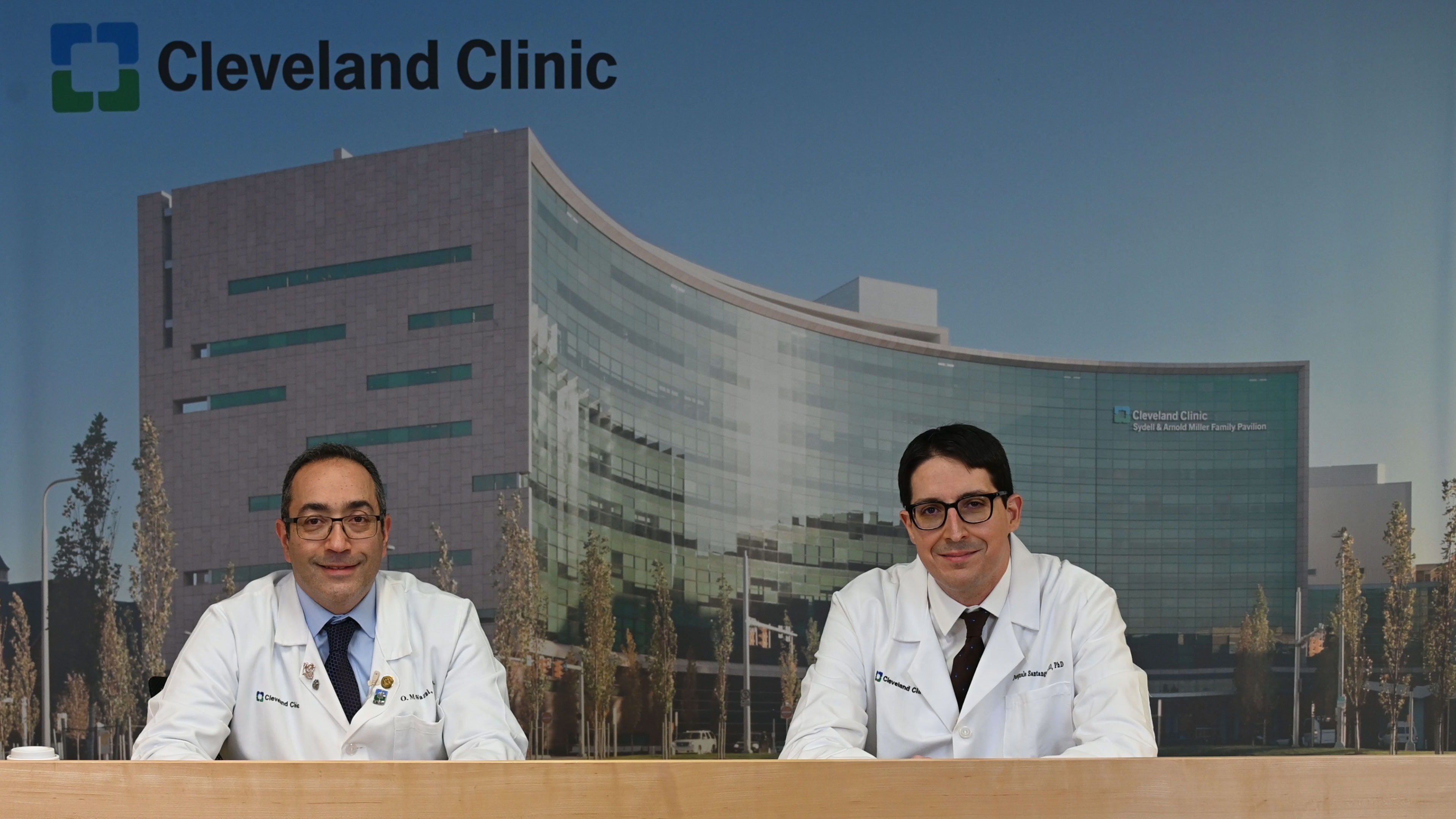
Oussama Wazni, MD, MBA, and Pasquale Santangeli, MD, PhD, from the Cardiac Electrophysiology and Pacing Section, discuss considerations for managing ventricular tachycardia as a result from cardiomyopathy.
Subscribe: Apple Podcasts | Buzzsprout | Spotify
Ventricular Tachycardia and Cardiomyopathy
Podcast Transcript
Announcer:
Welcome to Cleveland Clinic Cardiac Consult, brought to you by the Sydell and Arnold Miller Family Heart, Vascular and Thoracic Institute at Cleveland Clinic.
Oussama Wazni, MD, MBA:
Hello everyone, and welcome to another podcast from the EP section here at the Cleveland Clinic. I'm Oussama Wazni, I'm the section head, and today I'm joined by Dr. Pasquale Santangeli, our director for the VT Center. Welcome, Pasquale.
Pasquale Santangeli, MD, PhD:
Thank you very much.
Oussama Wazni, MD, MBA:
So today we're going to talk about a very interesting topic, which is management of ventricular tachycardia in both nonischemic cardiomyopathy and ischemic cardiomyopathy. Let's start with the difficult one first, which is nonischemic cardiomyopathy. Pasquale, tell us about your approach to ventricular tachycardia in a patient who has nonischemic cardiomyopathy.
Pasquale Santangeli, MD, PhD:
Right. It's a great question. Just in brief, the most important aspect, I think, is to understand the location of the substrate, essentially what we're dealing with. So really imaging comes into play here, is a very important aspect of it because really you can characterize the type of substrate, the dilation of the heart and the location of the scar essentially with light enhancements and so forth. And typically, they tend to fall into two peak categories, within the septal and anterior, we call the anteroseptal distribution, or the free wall in the left ventricle or right ventricle. And they have a lot of implications because if it is, for example, on the free wall, so not on the septum, tends to be epicardial more than endocardial. So, we do know upfront that we will most likely need to have an epicardial as well as an endocardial procedure. If it is septal, which is essentially mid-wall on the septum, we know that epicardial axis really doesn't have a lot of room there because there are coronary vessels and fat. And typically, we just approach them from the endocardium with typically radiofrequency ablation sequentially from the right side to left side of the septum. And when they fail, we use some form of bailout strategies, which we may talk about it later.
Oussama Wazni, MD, MBA:
Yeah, so when I started, I said the more difficult one. So, could you tell us why ablation of VT in nonischemic cardiomyopathy is actually more difficult?
Pasquale Santangeli, MD, PhD:
Yeah, the most difficult aspect of it is that some of the areas that are responsible for VT, some of the substrate is not accessible to us because with our mapping catheters it's just like a space rover. We just can touch the surface and we have to imagine what's happening underneath the surface in the mid-myocardial space. And this is true from the endocardium and epicardium. So, whenever you have an intramural substrate, for us, it's all about guessing where the scar would be or where the critical aspects of the VT will be. That's the challenging part. Now, assuming that we find where it's coming from, then there is a limitation of our ablation tools because radiofrequency can only penetrate up to about five millimeters on average and sometimes, we cannot reach these areas of interest.
Oussama Wazni, MD, MBA:
Now in the beginning you said imaging is very important and I would like to emphasize that we've actually made big strides in imaging with our Imaging colleagues and now we're to the point where we can use MRI imaging even in patients who have devices to characterize the substrate. So, any comments on that and how far have we gone and how much more do we have to go before we can really use those imaging techniques to the best abilities?
Pasquale Santangeli, MD, PhD:
Yes. So as part of our protocols here, essentially whenever we have a patient with any cardiomyopathy going for defibrillator, we always do imaging before getting the device because the images are perfect at that point. But assuming that most patients already have devices, as you said, the sequences have been improving over the years and now to a point that we can actually see most of the areas of interest essentially with MRI. And we've done studies now in thousands of patients demonstrating safety of MRI scanners even in patients with technically non-conditional devices. So, we can do that, and we have a system into place that can allow that essentially.
Oussama Wazni, MD, MBA:
I think you're routinely doing this right now.
Pasquale Santangeli, MD, PhD:
Yes, we do that routinely.
Oussama Wazni, MD, MBA:
And we're using some software to help guide us to where we need to ablate.
Pasquale Santangeli, MD, PhD:
Essentially, yes, once we have the images, then we reconstruct a 3D model of the heart essentially, where the scar and the distribution, so we can integrate that with our mapping system to facilitate the procedure. And by doing that, the procedural times have gone down about 50%. Essentially most procedures can be done between three and four hours for these cases. Most patients go home the next day. A select group of patients have gone also home the same day when the procedure is particularly short. So, it's completely changed.
Oussama Wazni, MD, MBA:
So, this also applies to the patients with ischemic cardiomyopathy. So, let's switch now a little bit to ischemic cardiomyopathy and the differences between somebody who has had now an MI versus nonischemic cardiomyopathy.
Pasquale Santangeli, MD, PhD:
Yes. The biggest difference is that with ischemic cardiomyopathy, there is no guessing. We know where the substrate is located. All it takes is an echocardiogram. We see the wall thinning, the area of akinesis, there is now of course not contracting. So, we already know what it is. And we also know that it's typically, it's a very thin area, so we can access it from the endocardium typically from the inside of the heart. So, by doing that, we understand where the substrate is located, we know how to target it, but mostly it being very superficial, we're very successful at ablating it and with radiofrequency we're very successful to up to 70-80 percent with a single procedure. And most patients can come off medications that can be potentially toxic like amiodarone, for example.
Oussama Wazni, MD, MBA:
So, there are three more things I want to talk about. First of all, when radiofrequency fails, how can we reach those difficult areas and how do we deal with that? The second one I want to talk about is in a patient who is unstable, how do we manage that? And the third one is the outcomes. So, let's start with the first one. So, in the procedure we're using radiofrequency, and this one is probably more so in nonischemic cardiomyopathy and we can't terminate the VT or the PVC. Is there anything now that we're using that can help us with that?
Pasquale Santangeli, MD, PhD:
Yes, so we have different types of bailout strategies we call it, that we've been implementing here at Cleveland Clinic. In fact, two thirds of patients reviewed that recently, they come here because they already failed radiofrequency ablation somewhere else. So, we are already ready for these types of bailout strategies. Most of the time the problem is, is that the ablation energy is not able to penetrate deep enough, so more than five millimeters. The way we approach that, we have different techniques. Typically, we try to cannulate micro vessels essentially from the venous system and occasionally also from the arterial system, but mostly from the venous system that is feeding that area essentially, that is close to that intramural scar. We can inject alcohol through that, and we use different techniques to do that, which has been very successful.
We're also equipped to do so-called bipolar ablation. So bipolar ablation means essentially ablation from both sides of an area, essentially to reach the center of it. There are special tools to do that. We also have very good experience with it. And whenever we cannot reach because of the extension of the substrate, it's very extensive, or if the patient is so sick, there is so much advance heart failure, we cannot really take them to the lab, we also now can provide radioablation, which is a noninvasive way of ablating the tissue, just similar to what we do with cancer essentially with Gamma Knife. So, we reconstruct what we mapped during the first procedure, we know where the target is, and we do radiation therapy noninvasively essentially to target that area. So, we have options for essentially the wide range of patients that failed radiofrequency ablation.
Oussama Wazni, MD, MBA:
Perfect. And now moving on to the patient who is unstable. And this brings us to our collaboration with our heart failure specialists and our surgeons. First of all, with the heart failure specialist to optimize the patient and then the surgeon to give us some backup hemodynamic support, but also sometimes to give us access to an area that we want to ablate. So, could you comment on both these aspects?
Pasquale Santangeli, MD, PhD:
I think this is crucial. Most patients that come at least to see me because of ventricular tachycardia and heart failure, I always recommend to establish heart failure care with us because we did look at this years ago and we realize that even if we do eliminate ventricular tachycardia, within five years, patients end up having advanced heart failure and need some form of other therapy. It can be interventional therapy, for example, sometimes valve correction and valve therapies or surgery or even LVAD or transplant. So, it's very important that you stay within the same system so we can follow them longitudinally with that.
So, before the procedure, typically we stratify patients with a risk code that essentially, we developed and validated externally so we know exactly what type of pathway we will follow. For patients that have advanced heart failure with very low ejection fraction, NVT, typically this triggers also a consult with heart failure. So, we can come up with a decision on how to optimize the patients before the procedure and whether we should use some additional strategies like mechanical hemodynamic support like, for example, an LVAD like Impella, which is percutaneous and temporary or even ECMO very rarely. By doing this in a very stepwise and structured way, we had very good outcomes even in the sickest patient population.
Oussama Wazni, MD, MBA:
Very good. And then there are certain instances where we actually ask the surgeons to give us access to an area so we can map it well, especially in somebody who's had previous surgery and we need to go to the surface of the ventricle. And then finally, I want to talk about our outcomes. I think we have made very big strides. You talked briefly about how now we have shortened the procedure time, which is very important because the shorter the time spent in a procedure under general anesthesia, the better the outcomes with respect to potential complications. Can you elaborate more on the success and also our outcomes with respect to potential complications in these patients?
Pasquale Santangeli, MD, PhD:
Yes. So absolute priority for ventricular arrhythmia ablation, but for everything we do in EP is to minimize complications. Ideally, we want to get to 0 percent and maybe at some point we'll get to that point, but we're close. And I think it's crucial because we realize that every time there is a complication, even if you manage to treat the VT successfully, then the long-term outcomes are worse than a patient that didn't have a complication. So, for us, the first priority is to minimize, eliminate. We have some techniques to do that. We implemented, for example, vascular access with ultrasound consistently. We minimize the number of access sites, for example, we just use typically venous access now and not arterial access for most patients. We have better and faster mapping ways and mapping tools so we can get our mapping done within an hour roughly, so we can spend the rest of the procedure to target the ablation part, which is the most important aspect. And typically, in three or four hours, most procedures can be actually successfully done, and patients do fairly well with it.
Oussama Wazni, MD, MBA:
Very good. And I'm happy to report that our success rates, like we discussed, in ischemic cardiomyopathy are 70 to 80 percent with no recurrence, at least at one year. Most of our patients, we can deescalate their antiarrhythmic drugs. Our complication rate is now below 5 percent, which is something to be really proud of, and thank you for your efforts with this, because the national average is more than 7percent and ours now has dropped to less than 5 percent. I think even with more effort, we can drop it even lower. So, thank you very much for your attention. Thank you, Pasquale, for being here. Thank you for leading our efforts in VT ablation and VT management in general, and we hope to see you in another podcast from EP at the Cleveland Clinic. Thank you.
Announcer:
Thank you for listening. We hope you enjoyed the podcast. We welcome your comments and feedback. Please contact us at heart@ccf.org. Like what you heard? Subscribe wherever you get your podcasts or listen at clevelandclinic.org/cardiacconsultpodcast.

Cardiac Consult
A Cleveland Clinic podcast exploring heart, vascular and thoracic topics of interest to healthcare providers: medical and surgical treatments, diagnostic testing, medical conditions, and research, technology and practice issues.


