A transesophageal echocardiogram (TEE) is an ultrasound test that uses sound waves to create pictures of your heart. Unlike other types of echo tests, a TEE creates pictures from inside your body. Your provider guides a thin, flexible tube down your esophagus. A TEE can diagnose many problems, including valve disease, tumors, clots and infections.
Advertisement
Cleveland Clinic is a non-profit academic medical center. Advertising on our site helps support our mission. We do not endorse non-Cleveland Clinic products or services. Policy
A transesophageal echocardiogram (TEE) is a test that uses sound waves to make detailed pictures of your heart and upper aorta. A TEE takes pictures from the inside of your body. While you’re sedated, your provider guides a small device down your throat. This makes TEE different from another type of echocardiogram test, called a transthoracic echo (TTE). A TTE is done outside your chest, and nothing goes into your body.
Advertisement
Cleveland Clinic is a non-profit academic medical center. Advertising on our site helps support our mission. We do not endorse non-Cleveland Clinic products or services. Policy
You may need a transesophageal echocardiogram if:
Providers typically do a transthoracic echo first because it’s less invasive. But they may skip that and go right to using a transesophageal echo if you have:
Advertisement
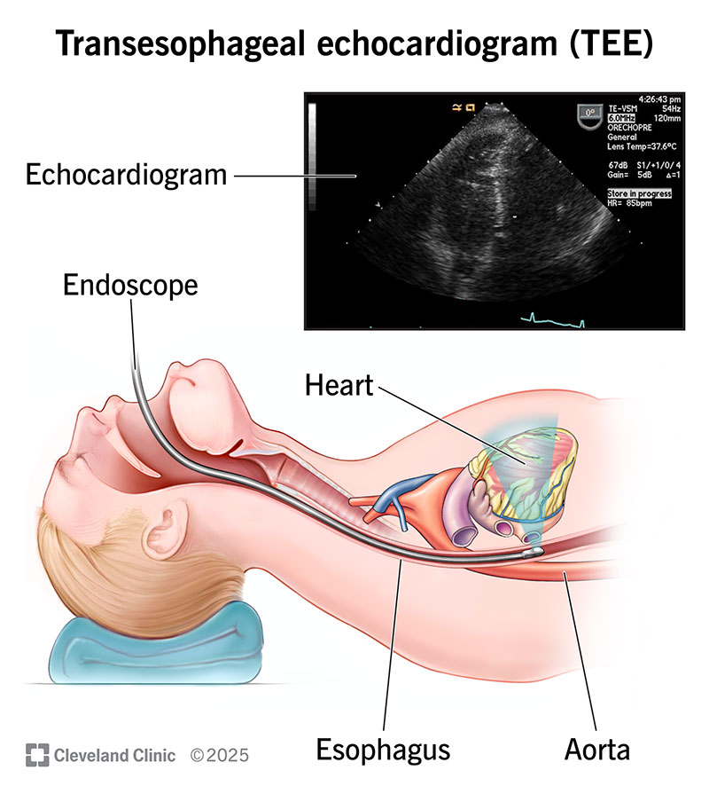
Image content: This image is available to view online.
View image online (https://my.clevelandclinic.org/-/scassets/images/org/health/articles/4992-tee-transesophageal-echocardiogram-illustration)
Your provider guides a long, thin tube called an endoscope down your esophagus. The tube has a transducer on its tip. This is a device that makes sound waves. These waves bounce off the different areas of your heart, causing echoes. The transducer sends these echoes to a computer that turns them into pictures. These pictures show the structure and function of your heart.
Your esophagus runs behind your heart. So, it’s the perfect spot for the transducer to send and receive sound waves. A TEE is especially valuable for examining structures in the back of your heart. These include your:
Your provider may combine TEE with Doppler ultrasound and color Doppler methods. These techniques show the speed and direction of blood flow through your heart.
Your provider will tell you how to prepare for your transesophageal echo. Their instructions will include:
As part of your prep, you’ll talk with your provider about your health history. Some conditions make a TEE riskier to do. Tell your provider if you have:
These conditions don’t necessarily make the TEE too risky to perform, but your provider should know if these conditions exist. Also, tell your provider if you take medication for:
To perform a transesophageal echocardiogram, your provider will:
Advertisement
A TEE may take up to 90 minutes. The picture-taking part usually only takes 15 minutes.
Possible complications include:
Talk with your provider about the risks of a TEE before your procedure.
Someone will need to drive you home from your TEE. You won’t be able to drive for 24 hours after the test due to the sedation. You can expect to:
Your provider will tell you when the test results will be available.
A transesophageal echocardiogram shows a detailed view of your heart’s structure and function. The results may help your provider diagnose a wide range of conditions, including:
Advertisement
You’re awake but drowsy. Your provider will sedate you to make you feel more comfortable during the test. Most people don’t need general anesthesia (deep sleep). If you do receive it, you’ll be fully asleep and won’t remember the procedure. But recovery may take longer compared with moderate sedation.
No. It’s a diagnostic test that checks your heart. A TEE is an invasive procedure because your provider needs to put a device inside your body, but it’s not surgery. You may experience mild discomfort. But the risk of serious problems is low.
A device going down your throat probably doesn’t sound too pleasant. Your provider knows you might feel a bit anxious about getting a transesophageal echo. But they’ll help you stay as comfortable as possible. They’ll give you medicine to help you relax and avoid feeling pain.
Just remember, this test can be lifesaving. And it’ll be over before you know it. Ask your provider if you have any questions about how to prepare or what the results may show.
Advertisement


Sign up for our Health Essentials emails for expert guidance on nutrition, fitness, sleep, skin care and more.
Learn more about the Health Library and our editorial process.
Cleveland Clinic’s health articles are based on evidence-backed information and review by medical professionals to ensure accuracy, reliability and up-to-date clinical standards.
Cleveland Clinic’s health articles are based on evidence-backed information and review by medical professionals to ensure accuracy, reliability and up-to-date clinical standards.
When your heart needs some help, the cardiology experts at Cleveland Clinic are here for you. We diagnose and treat the full spectrum of cardiovascular diseases.
