The 20-week ultrasound scan, sometimes called an anatomy or anomaly scan, happens around 18 to 22 weeks of pregnancy. It checks the development of fetal organs and body parts and can detect certain congenital conditions. In most cases, you can also learn the sex of the fetus.
Advertisement
Cleveland Clinic is a non-profit academic medical center. Advertising on our site helps support our mission. We do not endorse non-Cleveland Clinic products or services. Policy
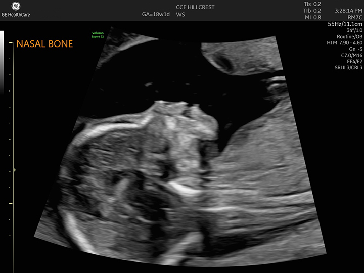
Image content: This image is available to view online.
View image online (https://my.clevelandclinic.org/-/scassets/images/org/health/articles/22644-20-week-ultrasound)
A 20-week ultrasound is a prenatal ultrasound that happens between 18 and 22 weeks of pregnancy. It’s a way to check on how the fetus is growing. It also looks for signs of congenital conditions or how an organ or body part looks.
Advertisement
Cleveland Clinic is a non-profit academic medical center. Advertising on our site helps support our mission. We do not endorse non-Cleveland Clinic products or services. Policy
The scan is one tool your pregnancy care provider uses to make sure the fetus and pregnancy are healthy. It also helps them catch any potential problems before birth. A 20-week ultrasound is also called an anatomy scan.
During your appointment, an ultrasound technician (sonographer) takes pictures and measurements of the fetal:
They also:
Ultrasounds use sound waves to make pictures of the inside of your body. The technician will move a device called a transducer across your belly to get the images they need of the fetus. They’ll also take certain measurements and look at organs and body parts. Your provider uses the images to check on how the fetus is growing and look for possible health issues.
There isn’t much to do to prepare for your appointment. This will be one of your longer prenatal ultrasound appointments — around 45 minutes. This will likely be a separate appointment from your regular pregnancy care visit. Some providers suggest eating before. This may make the fetus move more, which can help the technician get all the measurements they need.
Advertisement
Here is what you can expect:
Generally, your ultrasound technician will be quiet as they work. Don’t be alarmed if they don’t talk to you or share results as they go. Your obstetrician (or other healthcare provider) will go over the results with you either immediately after or at a follow-up appointment in the next few days.
Yes. Depending on the position the fetus is in, your technician may be able to see its genitals. If you don’t want to know the sex, speak up so they don’t accidentally spoil the surprise.
A 20-week ultrasound isn’t dangerous for you or the fetus. There aren’t harmful risks to the scan, but there’s a chance you could get false results.
The benefits outweigh any drawbacks, though. The ultrasound helps detect potentially life-altering health conditions. This can allow both you and your healthcare team to plan for any extra care your baby may need after birth.
The test tells you and your pregnancy care team if the fetus is developing at a healthy rate for its age. It also checks for signs of congenital conditions (based on your chromosomes, or family history) and possible structural issues with major organs like its brain, heart and kidneys. Your provider also looks closely at the placenta’s position and checks to make sure there’s a safe amount of amniotic fluid.
The 20-week ultrasound doesn’t diagnose anything, though. If your pregnancy care provider is concerned about anything they see, they’ll order more tests.
Every facility does things a little differently, but in general, you’ll probably talk to your obstetrician right after the scan. The ultrasound tech will pass along the images and their findings, and then your provider will go over the results with you. The anatomy scan is usually a positive and reassuring experience. Most of the time, no major issues are found.
Advertisement
A 20-week ultrasound doesn’t find all congenital conditions. But the scan can help detect several serious conditions, like:
If your healthcare provider has concerns about anything in your 20-week scan, they may recommend:
Speak with your healthcare provider early in your pregnancy to make sure you understand the types of noninvasive prenatal testing available to you. Some tests can happen before your 20-week ultrasound.
It’s normal to have a lot of questions at your 20-week ultrasound. Some of the questions you may want to ask include:
It’s normal to be both excited and nervous about your 20-week ultrasound appointment. You’ll get to see the fetus and find out how it’s developing. Talk to your healthcare provider about any concerns you have so they can offer reassurance and ease your worries. In most cases, the 20-week anatomy scan is a positive experience for soon-to-be parents.
Advertisement


Sign up for our Health Essentials emails for expert guidance on nutrition, fitness, sleep, skin care and more.
Learn more about the Health Library and our editorial process.
Cleveland Clinic’s health articles are based on evidence-backed information and review by medical professionals to ensure accuracy, reliability and up-to-date clinical standards.
Cleveland Clinic’s health articles are based on evidence-backed information and review by medical professionals to ensure accuracy, reliability and up-to-date clinical standards.
Prenatal tests can give your providers information about your pregnancy and fetal development. Cleveland Clinic’s experts can guide you through prenatal testing.
