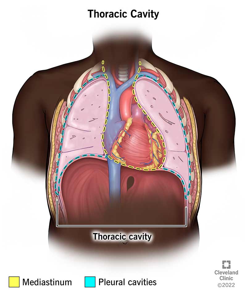
Your thoracic cavity is a space in your chest that contains organs, blood vessels, nerves and other important body structures. It’s divided into three main parts: right pleural cavity, left pleural cavity and mediastinum. The five organs in your thoracic cavity are your heart, lungs, esophagus, trachea and thymus.
Advertisement
Cleveland Clinic is a non-profit academic medical center. Advertising on our site helps support our mission. We do not endorse non-Cleveland Clinic products or services. Policy
Your thoracic cavity (chest cavity) is a space inside your thorax (chest) that contains your heart, lungs and other organs and tissues. It’s the second biggest hollow space in your body, with only your abdominal cavity being larger.
Advertisement
Cleveland Clinic is a non-profit academic medical center. Advertising on our site helps support our mission. We do not endorse non-Cleveland Clinic products or services. Policy
Your thoracic cavity houses the organs and tissues in your chest. These organs and tissues play a vital role in many of your body’s systems, including your:
Your thoracic cavity contains five organs:

Image content: This image is available to view online.
View image online (https://my.clevelandclinic.org/-/scassets/images/org/health/articles/24748-thoracic-cavity-illustration.jpg)
Your thoracic cavity is located in your chest. It’s enclosed by the bones and muscles that make up your chest wall. Your thoracic cavity begins just below your neck and ends at the bottom of your ribcage.
Here’s a bit more detail about your thoracic cavity’s boundaries at different locations within your chest:
Advertisement
Your thoracic cavity is like a home with many rooms. There are three main “rooms” in your thoracic cavity:
Your mediastinum is further divided into several parts. They’re named according to their position in your chest. There are two main classification systems that scientists use. The first is an older system that divides the mediastinum into four parts:
A more recent classification system divides the mediastinum into three parts with different names:
The older model is based on X-ray images of your heart. The newer model is based on cross-sectional imaging (like CT scans). So, the newer model is helpful for healthcare providers who use cross-sectional imaging for diagnosis and treatment.
Your thoracic cavity includes organs, blood vessels, nerves and connective tissues. Some key components include:
A thin membrane called the pleura lines your thoracic cavity. This layer of tissue helps protect the organs and structures inside your thoracic cavity.
A wide range of conditions and disorders can affect the organs and tissues within your thoracic cavity. These include:
There are many tests your healthcare provider may use to check the health of organs and tissues in your thoracic cavity, including:
There’s a lot you can do to keep the organs and tissues in your thoracic cavity healthy. Here are some tips:
Advertisement
Your thoracic cavity is the large space in your chest where some of your body’s most important work gets done. If you’re interested in learning about the thoracic cavity, you may have had a recent lung or heart disease diagnosis. Or maybe you just want to know more about the human body. Either way, your healthcare provider can help you learn more. Ask them to share resources or help explain anything that you find confusing as you learn about the inner workings of your body.
Advertisement

Sign up for our Health Essentials emails for expert guidance on nutrition, fitness, sleep, skin care and more.
Learn more about the Health Library and our editorial process.
Cleveland Clinic’s health articles are based on evidence-backed information and review by medical professionals to ensure accuracy, reliability and up-to-date clinical standards.
Cleveland Clinic’s health articles are based on evidence-backed information and review by medical professionals to ensure accuracy, reliability and up-to-date clinical standards.