Your thoracic duct is part of your lymphatic system. It runs vertically through the middle of your chest, near your spine. It drains lymph from many different areas of your body and empties it into large veins in your upper chest. Traumatic injuries and surgery complications can damage your thoracic duct, causing fluid leaks (these are treatable).
Advertisement
Cleveland Clinic is a non-profit academic medical center. Advertising on our site helps support our mission. We do not endorse non-Cleveland Clinic products or services. Policy
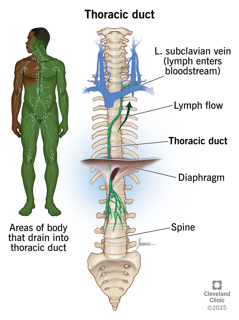
Image content: This image is available to view online.
View image online (https://my.clevelandclinic.org/-/scassets/images/org/health/articles/thoracic-duct)
Your thoracic duct is the biggest lymphatic vessel in your body. It’s a long, tubelike structure that carries lymph from your belly area up through your chest. Like a highway on-ramp, your thoracic duct merges into large veins near the base of your neck. This allows lymph to rejoin your bloodstream and continue its journey through your body.
Advertisement
Cleveland Clinic is a non-profit academic medical center. Advertising on our site helps support our mission. We do not endorse non-Cleveland Clinic products or services. Policy
Your thoracic duct and another duct, called your right lymphatic duct, are the two main pathways that help lymph return to your bloodstream. Your right lymphatic duct drains lymph from your right arm and the right sides of your head, neck and chest. Your thoracic duct drains lymph from all the other parts of your body, including:
Your thoracic duct collects lymph from many smaller lymphatic vessels throughout your body. It delivers this lymph to your circulatory system. This helps support healthy fluid levels and allows your body to filter out waste.
Here’s what that process looks like:
Advertisement
Each day, your thoracic duct delivers about 3 liters of lymph into your circulatory system. This includes chyle, a special kind of lymph that’s rich in fats (lipids). Chyle comes from your digestive system. It’s milky-white in color, unlike lymph from elsewhere in your body, which is typically clear.
Your thoracic duct is in the middle of your chest. It’s close to your aorta, esophagus and spine. It starts near the top of your lumbar spine (between the T12 and L2 vertebrae in most people). That’s where several lymphatic vessels merge — including those that carry lymph from your legs and intestines. These vessels sometimes form a sac-like structure called the cisterna chyli.
From there, your thoracic duct travels upward. It follows a similar course to your spine as it travels through your chest (thorax). Along the way, other lymphatic vessels drain into it. As it nears your neck, it forms an arch that rises just above your collarbone. It then curves back downward to empty into one or more veins in your upper chest.
The exact drainage location varies from person to person. Your thoracic duct might drain into the spot where your left subclavian vein and left internal jugular vein meet. Or it might drain into either of these veins, very close to their junction. It’s also possible for it to drain into your left external jugular vein.
Your thoracic duct may lead directly to a vein. This is the case for most people. But there are variations. Your thoracic duct might split into branches before coming back together to form a single terminal (end-point) duct. Or it could split into two ducts that open separately into your veins.
You might not need to know the exact anatomy of your thoracic duct. But healthcare providers keep it in mind when doing certain procedures and surgeries.
Your thoracic duct is shaped like a tube. It has many other tubes — smaller lymphatic vessels — connected to it. The walls of your thoracic duct have three layers. These are called the intima, media and adventitia. Smooth muscle tissue in the middle layer (media) squeezes regularly. This keeps lymph moving in the right direction.
As lymph flows through the duct, it encounters valves at various points. These valves open and close to further help lymph along on its journey.
Your thoracic duct is anywhere from 38 to 45 centimeters long. Its opening (diameter) varies along its length from 2 to 5 centimeters.
Conditions that may keep your thoracic duct from working properly include:
Advertisement
Treatment depends on the condition but may include:
Your healthcare provider can tell you more about what to expect in your situation.
If you’re having surgery on your chest or belly, your provider will tell you if thoracic duct injury is a possible complication. They’ll also tell you what signs to look out for as you recover.
Your thoracic duct is one of many body parts that work quietly behind the scenes, helping your body function from day to day. If you wanted to point to your thoracic duct, you’d have to trace a line from your belly to your upper chest. It’s a long tube that does important work for your lymphatic system.
Because it follows such a long course, there are more chances for your thoracic duct to get injured. Still, such injuries aren’t common. And your provider is prepared to treat them if they happen.
Advertisement


Sign up for our Health Essentials emails for expert guidance on nutrition, fitness, sleep, skin care and more.
Learn more about the Health Library and our editorial process.
Cleveland Clinic’s health articles are based on evidence-backed information and review by medical professionals to ensure accuracy, reliability and up-to-date clinical standards.
Cleveland Clinic’s health articles are based on evidence-backed information and review by medical professionals to ensure accuracy, reliability and up-to-date clinical standards.
Cleveland Clinic’s primary care providers offer lifelong medical care. From sinus infections and high blood pressure to preventive screening, we’re here for you.
