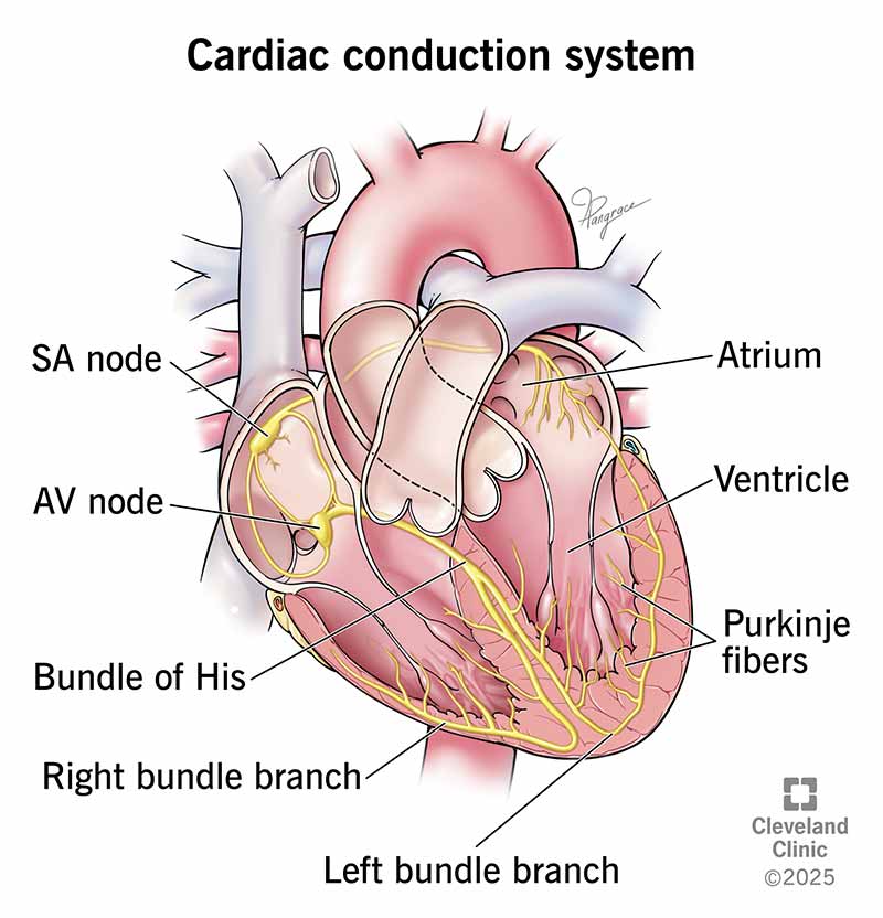
Your cardiac conduction system is the network of nodes, cells and signals that controls your heartbeat. Each time your heart beats, electrical signals travel through your heart. These signals cause different parts of your heart to expand and contract. These actions regulate blood flow through your heart and body.
Advertisement
Cleveland Clinic is a non-profit academic medical center. Advertising on our site helps support our mission. We do not endorse non-Cleveland Clinic products or services. Policy
Your heart’s conduction system is the network of nodes (groups of cells that can be either nerve or muscle tissue), specialized cells and electrical signals that keep your heart beating.
Advertisement
Cleveland Clinic is a non-profit academic medical center. Advertising on our site helps support our mission. We do not endorse non-Cleveland Clinic products or services. Policy
Two types of cells control your heartbeat:
Signals tell your heart when to pump blood through your body.
Your heart (cardiac) conduction system sends the signal to start a heartbeat. It also sends signals that tell different parts of your heart to relax and contract (squeeze). This process of contracting and relaxing controls blood flow through your heart and to the rest of your body.
Ideally, the electrical conduction system keeps up a steady, even heart rate. It also helps your heart speed up when you need more blood and oxygen or slow down when it’s time to rest.
For each heartbeat, electrical signals travel through the conduction pathway of your heart. It starts when your sinoatrial (SA) node creates an excitation signal. This electrical signal is like electricity traveling through wires to an appliance in your home.
The excitation signal travels to:
Advertisement
These steps make up one full contraction of your heart muscle. Your heart’s electrical conduction system sends out thousands of signals per day to keep your heart beating.

Image content: This image is available to view online.
View image online (https://my.clevelandclinic.org/-/scassets/images/org/health/articles/heart-conduction-system)
The conduction system in your heart contains specialized cells and nodes that control your heartbeat. These are the:
Your sinoatrial (SA) node is your heart’s natural pacemaker. It sends the electrical impulses that start your heartbeat. When your sinoatrial node isn’t working well, the lower segments of your conduction system act as backup pacemaker cells.
The SA node is in the upper part of your heart’s right atrium. It’s at the edge of your atrium near your superior vena cava (a large vein that brings oxygen-poor blood from your body to your heart).
Your autonomic nervous system controls how quickly or slowly your SA node sends electrical signals. This part of your nervous system directs hormones that control your heart rate based on what you’re doing. For example, your heart rate increases during physical activity and slows when you’re asleep.
The autonomic nervous system includes your:
Your atrioventricular (AV) node, located near the central area of your heart, delays the SA node’s electrical signal. It delays the signal by a consistent amount of time (a fraction of a second) each time.
The delay ensures that your atria (upper heart chambers) are empty before the contraction stops. Your atria receive blood from your body and empty it into your ventricles (lower heart chambers).
The bundle of His (sounds like “hiss”) is a branch of fibers (nerve cells) that extends from your AV node. This fiber bundle receives the electrical signal from the AV node and carries it to the Purkinje fibers.
The bundle of His runs down the length of the septum (wall) that separates your right and left ventricles. The atrioventricular bundle has two branches:
The Purkinje fibers are branches of specialized nerve cells. They send electrical signals very quickly to your heart’s right and left ventricles. Purkinje fibers are in your ventricle walls in the inner layer of tissue that lines your heart’s chambers.
Advertisement
When the Purkinje fibers deliver electrical signals to your ventricles, the ventricles contract. Blood flows from your right ventricle to your pulmonary arteries and from your left ventricle to your aorta. Your pulmonary arteries take blood to your lungs to get oxygen. Your aorta, your body’s largest artery, sends blood from your heart to the rest of your body.
The sinoatrial node is about 15 millimeters (mm) long (about as long as a staple) and 4 mm wide. It looks like a key, with a head and a smaller, jagged lower part. The AV node is about 5 mm long and 5 mm wide. This is about the size of a light switch tip, but the AV node looks like a spindle. The Bundle of His is 20 mm long and about 4 mm around.
Together, the Bundle of His, its branches and the Purkinje fibers look like an upside-down tree. The Bundle of His and its branches are like a tree trunk. The Purkinje fibers spread upward and outward to look like a tree’s canopy.
Several different conditions can affect your heart’s electrical system. Cardiac conduction problems cause issues with your heart’s rhythm.
Some common heart rhythm disorders include:
Advertisement
If you have an issue with your cardiac conduction system, you may experience:
You should seek emergency medical care if you have these symptoms. A healthcare provider can use an electrocardiogram (EKG) to check your heart rhythm. If they want to look at your heart’s electrical activity longer, your provider may ask you to wear a heart monitor for a few days or weeks. An electrophysiology study can give even more information about your cardiac conduction system.
Medicines and procedures like a pacemaker placement can treat issues with your cardiac electrical system.
You can’t change the genetics that cause many problems with heart rhythm. But you can help keep your entire heart well with healthy habits. You can:
You may not be aware of your heart’s electrical system until you have an issue with your heart’s rhythm. Conduction signals keep your heart beating, which moves blood through your heart. Knowing how this system works can help you spot potential problems and seek help. You can keep your heart’s conduction system and your entire heart healthy by managing stress and being physically active.
Advertisement

Sign up for our Health Essentials emails for expert guidance on nutrition, fitness, sleep, skin care and more.
Learn more about the Health Library and our editorial process.
Cleveland Clinic’s health articles are based on evidence-backed information and review by medical professionals to ensure accuracy, reliability and up-to-date clinical standards.
Cleveland Clinic’s health articles are based on evidence-backed information and review by medical professionals to ensure accuracy, reliability and up-to-date clinical standards.
When your heart needs some help, the cardiology experts at Cleveland Clinic are here for you. We diagnose and treat the full spectrum of cardiovascular diseases.
