An echocardiogram (also called an echo or heart ultrasound) is a test that checks your heart’s structure and function. An echo can diagnose many heart problems, including cardiomyopathy and valve disease. There are several types of echo tests, including transthoracic and transesophageal. Talk with your provider about the type that’s best for you.
Advertisement
Cleveland Clinic is a non-profit academic medical center. Advertising on our site helps support our mission. We do not endorse non-Cleveland Clinic products or services. Policy
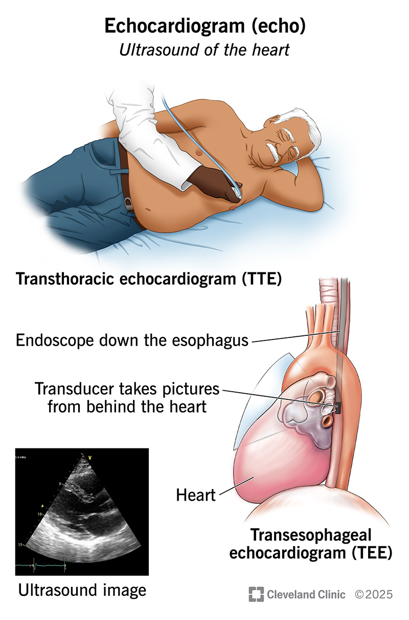
Image content: This image is available to view online.
View image online (https://my.clevelandclinic.org/-/scassets/images/org/health/articles/16947-echocardiogram)
An echocardiogram is an ultrasound test for your heart. It uses high-frequency sound waves to create moving pictures of your heart as it beats. This test can help your healthcare provider diagnose and treat many different heart conditions, including those you’re born with and those you develop later in life. Other names for this test include echo, heart echo and heart/cardiac ultrasound.
Advertisement
Cleveland Clinic is a non-profit academic medical center. Advertising on our site helps support our mission. We do not endorse non-Cleveland Clinic products or services. Policy
An echocardiogram shows your heart’s structure and pumping ability. It also shows blood flow through your heart, including through each of its valves.
Cardiologists order echocardiograms for many reasons, including to:
Your provider will tell you why you need this test and how it can help you. Most echocardiograms are noninvasive, meaning nothing goes into your body. In some cases, you may need a more invasive type where a small device takes pictures from inside your body.
There are several types of echo tests:
Advertisement
An echocardiogram uses ultrasound technology to create images of your heart in motion. A device called a transducer sends sound waves through your chest. These waves bounce off your heart tissue, making echoes. The transducer captures these echoes and sends them back to a computer.
Computer software uses these echoes to make pictures of your heart. The pictures move, allowing your healthcare provider to see how your heart beats. The pictures are easily saved for your provider to review and keep as part of your medical record.
This same basic technology is the same for all echocardiograms. But providers may use certain techniques to get extra details or check a specific aspect of your heart, like blood flow through your valves. Examples of such techniques include:
An echo doesn’t use radiation, which many people see as a benefit. This is different from tests like X-rays and CT scans, which use small amounts of radiation.
Prep depends on the type of echo you’re getting. Your provider will give you specific instructions. You may need to:
For most types of echo tests, you’ll need to undress from the waist up and put on a hospital gown.
A cardiac sonographer performs your echo. The test usually takes 40 to 60 minutes. A TEE may take up to 90 minutes. The exact details vary by type. In general, your provider will:
Advertisement
Once your provider has enough pictures, they’ll wrap up the test. If you were in a gown, you’ll change back into your clothes. You may need to stay for around 30 minutes so your provider can monitor you after the test.
Echocardiography is typically safe and painless. A TTE has no known risks. Other types, like TEE and those that stress your heart, come with some risks. For example, an exercise stress test may make you feel symptoms like chest pain or shortness of breath. A TEE may cause a mild sore throat for a few days. The contrast dye used in some echo tests may cause an allergic reaction.
Your provider will explain the risks before your test and answer all your questions.
For most echo tests, you can drive yourself home. But you need someone to drive you after a TEE because you’re sedated during this test. It’s not safe for you to drive for 24 hours.
Your provider who ordered the test (likely your heart doctor) will let you know when the results are ready. You may have a follow-up visit or phone call to discuss them.
You might see results pop up on your electronic medical record. But it’s a good idea to wait for your provider to review them with you. Certain things may sound scary when you read the results on your own. Your provider will help you understand what the results mean and whether you need treatment for any issues.
Advertisement
An echo makes moving pictures of your heart that your provider can review. They may show you these pictures when you come in to discuss the results. A normal echocardiogram means there aren’t any concerns about your heart’s structure or function. An echo can also find problems that need treatment either right now or down the road.
An echocardiogram can diagnose:
Call the doctor who ordered your echo if you have:
An echocardiogram is a common test that can reveal a lot about your heart’s structure and function. If your provider recommends an echo for you, ask about the type you’ll receive and what you can expect.
Advertisement
It’s common to feel a bit anxious before medical tests. If you do, it’s OK to tell the sonographer who’s doing your echo. They’ll help you relax and feel as comfortable as possible. It’ll be over before you know it. Just remember, you’re getting valuable information to help take good care of your heart.


Sign up for our Health Essentials emails for expert guidance on nutrition, fitness, sleep, skin care and more.
Learn more about the Health Library and our editorial process.
Cleveland Clinic’s health articles are based on evidence-backed information and review by medical professionals to ensure accuracy, reliability and up-to-date clinical standards.
Cleveland Clinic’s health articles are based on evidence-backed information and review by medical professionals to ensure accuracy, reliability and up-to-date clinical standards.
When your heart needs some help, the cardiology experts at Cleveland Clinic are here for you. We diagnose and treat the full spectrum of cardiovascular diseases.
