The First Stopped Heart Surgery

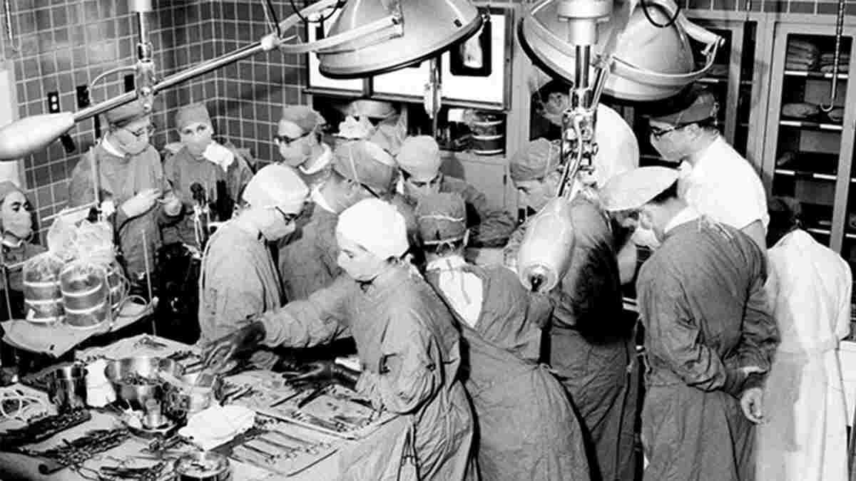
The first documented stopped heart surgery was performed on February 17, 1956. Dr. Lars Svensson, Chairman of the Heart, Vascular and Thoracic Institute reviews the operative notes from this innovative surgery performed by Dr. Donald Effler and Dr. Lawrence Groves, using a heart lung machine developed by Dr. Willem Kolff at Cleveland Clinic. Dr. Svensson uses illustrations to take us step by step through the procedure. The majority of heart procedures done today use a similar process to stop the heart as the one that Drs. Effler and Groves used 65 years ago during this ground-breaking surgery.
Watch Dr. Svensson illustrate this surgery step by step
Subscribe: Apple Podcasts | Buzzsprout | Spotify
The First Stopped Heart Surgery
Podcast Transcript
Announcer:
Welcome to Love Your Heart, brought to you by Cleveland Clinic's Sydell and Arnold Miller Family Heart, Vascular and Thoracic Institute. These podcasts will help you learn more about your heart, thoracic and vascular systems, ways to stay healthy and information about diseases and treatment options. Enjoy.
Lars Svensson, MD, PhD:
I'm Lars Svensson, Chairman of the Heart, Vascular and Thoracic Institute. And it had always been an interest to me, the so-called first stopped heart at the Cleveland Clinic. Now, many of you know about the story of Sones and flipping the catheter into the right coronary artery, and showing a catheterization. And also the early pioneering work of Favaloro on coronary artery bypass surgery. But there was something that was fascinating about the story at the Cleveland Clinic of the first stopped heart. So I decided to see if I could find out more about it. At the same time we interviewed the patient who was involved in this operation. Turned out this operation was done before the first cardiac catheterization, and actually a long time before, also the first coronary artery bypass operations.
Lars Svensson, MD, PhD:
So we got hold of the micro fiches from deep in the dungeons of the store rooms, and they then sent me what they could extract from these micro fiches. This is something that some of you may remember, was basically a photograph taken of charts. And then you looked under an enlargement screen at it. Well, needless to say, the handwriting was pretty bad, and it was faded because this was written in pen, and the pen writing had faded. But I happen to have very bad handwriting myself. So it takes a bit of an abstract idea to figure out what the writing said. I wouldn't say this is a Rosetta Stone, but it was trying to guess what was written. And I managed to figure out quite a lot about it. And then I dug up further research about this particular patient.
Lars Svensson, MD, PhD:
So 65 years ago in 1956, in February of that year, on the 17th, this patient underwent this operation. He was in heart failure. He'd been diagnosed as having repeated bouts of pneumonia, but it was actually pulmonary edema, and his lungs were flooding the water. He was only 17 months old. And at that time Dr. Effler had been doing research on trying to stop the heart on the heart lung-machine in patients. And this was the first child that they decided to try it on. So Dr. Sones, before he famous for his catheterization, spoke to the family and said, "Dr. Effler thinks he can do this operation, but the risk of death is going to be, probably 25 to 30%." So the family agreed, because nothing was getting better. They were having to carry their child around the whole time, child couldn't sleep, was in pulmonary edema all the time. And they agreed to go ahead.
Lars Svensson, MD, PhD:
So the operation was done. And essentially what they did, they put the patient on a heart-lung machine that had been developed here at the Cleveland Clinic by Dr. Kolff, and Dr. Groves was the assistant. They put in a, what we call arterial line. So this is the oxygenated blood going into an artery, they happened to use the subclavian artery, which was also the first time, really that this was used as a site of perfusion. We actually have shown in some big series here at the Cleveland Clinic, it's a very safe way to put patients on the heart-lung machine. And in fact reduces the risk of stroke by some 40% in some patients.
Lars Svensson, MD, PhD:
Then what they did was they drained the blood out of the superior and vena cava. And then they put these loops, as we call them. So basically strings around the superior and vena cava, isolated the heart. And that allowed them then to put the patient on the heart-lung machine, snare up these tapes, and then they put a clamp on the aorta, and injected a higher concentration potassium solution into what we call the aortic root. So just about the aortic valve, and that perfused the coronary arteries and stopped the heart. Potassium stops the heart. And then they were able to do the operation.
Lars Svensson, MD, PhD:
Why is this so important? Well, in the United States, there's somewhere around half a million, 500,000, heart operations done every year. And the majority of those, probably 80%, are done with a stopped heart with potassium stopping the heart. And then as soon as you reperfuse the heart, as we call it, take the clamp off, let the blood flow to the coronary arteries again. The heart starts beating again. Sometimes we have to give it a bit of a zap, a little shock to get it back into rhythm, but it's amazingly safe. And here at the Cleveland Clinic, for all our heart operations, we run in a region of a 1.3 to 1.7 annual risk of death, with some 4,500 heart operations we do every year. Here at main campus, for our whole system, it's several thousands that we operate every year.
Lars Svensson, MD, PhD:
So this has become the preferred technique for doing a heart operations. And that was due to the pioneering work of Dr. Effler, Dr. Groves and the encouragement of Dr. Sones. So what I'm going to do now, is just draw and take some of the original figures that were made by Dr. Effler and Dr. Groves for the article on the first three patients they did, and try and illustrate for you, what they actually did.
Lars Svensson, MD, PhD:
Alrighty. Here as promised is the diagram of the first stopped heart operation. So this is cardiac surgery 101 in more ways than one, because this is how the first heart operation was done with a stopped heart and the way we do it nowadays. So let me show you first, a couple of things here. So the heart has two pumping chambers, the filling chamber up here, the right atrium, and then the left atrium is up there. Then you have the right pumping chamber and the left pumping chamber. And in this patient, there was a hole over here, with blood leaking across what we call the septum, into the right ventricle from the left ventricle. So instead of the blood going out into the aorta that way, it was going into the right ventricle. And then from there into the pulmonary arteries and flooding the lungs with fluid.
Lars Svensson, MD, PhD:
And essentially it's a bit like the child was drowning in the extra high blood pressure that the child had in the lungs. So the problem was then to try and fix this hole over here. So what they did was, they put, first of all, what we call a arterial cannula into the left subclavian artery. And we use these arteries, as I said earlier, for putting a lot of patients on the heart lung machines with complex problems, particularly aortic aneurysms and reoperations. So the blood comes in here, it has oxygen in it, it goes down to the body, into what we call the innominate artery, which goes to the right side of the brain, and to the left side of the brain, the carotid. So that's blood coming in, and also for the heart, it comes to the coronary arteries, coming off here and over here.
Lars Svensson, MD, PhD:
Then to get the oxygen into the blood, they put a cannula through the right atrium. So the right filling chamber, up into the superior vena cava, and then they put another one into the inferior vena cava. And as I said, they put these loops, or tapes, around the vena cava, so they could isolate the heart for the operation. So that's the first part of the preparation. Then what they did was, they put these snares down. And what that does, is it stops blood coming into the heart. So in other words, the blood is now being drained out into this cannula, out into this cannula, into the cardiopulmonary bypass circuit, as we call it. And then back into the aorta there to give blood to the rest of the body.
Lars Svensson, MD, PhD:
Then what they did was they put a clamp across the aorta, so they isolated there. And then they injected this high concentration potassium solution, potassium citrate, into the aortic root and that stopped the heart. So now they had isolated the heart, they had stopped the heart, and then the next step was then to do the repair. So we'll flip over to the next one to show you that part of the operation. So what they did was, now that they had isolated the heart, they went through the right ventricle, and cut a hole in the right ventricle. And then they could see a hole in the heart, what we call a ventricular septal defect. And they then stitched that up with these stitches as you see there, closed the hole, and then they just have to stitch up the right ventricle again.
Lars Svensson, MD, PhD:
We do it slightly differently nowadays, quite often in kids, we will come through the tricuspid valve and repair the hole in the septum, or sometimes we'll even come in through the aortic root, and work through the aortic valve. Because this does somewhat damage to the right ventricle, going through it. So we try and avoid that as much as possible.
Lars Svensson, MD, PhD:
After the operation, the patient came back to see Dr. Effler again. And he said, "Your lucky number is 17. And the reason is you were 17 months old. You were operated on the 17th of February, you weighed 17 pounds, and we stopped your heart on the heart-lung machine for 17 minutes to do the operation." That in itself was quite an achievement in those days. And this was the beginning of heart surgery on the heart lung machine at the Cleveland Clinic that we do every day. Tomorrow, we have 21 patients on for heart surgery, and most of those are going to be done with a stopped heart. Thank you for watching this little interesting vignette about the stopped heart at the Cleveland Clinic. That set the stage for all heart surgery around the world. Thank you.
Announcer:
Thank you for listening. We hope you enjoyed the podcast. We welcome your comments and feedback. Please contact us at heart@ccf.org. Like what you heard? Subscribe wherever you get your podcasts, or listen at clevelandclinic.org/loveyourheartpodcast.

Love Your Heart
A Cleveland Clinic podcast to help you learn more about heart and vascular disease and conditions affecting your chest. We explore prevention, diagnostic tests, medical and surgical treatments, new innovations and more.


