Characteristics and Long Term Outcomes in Adults with Marfan Syndrome and Ascending Aorta Surgery

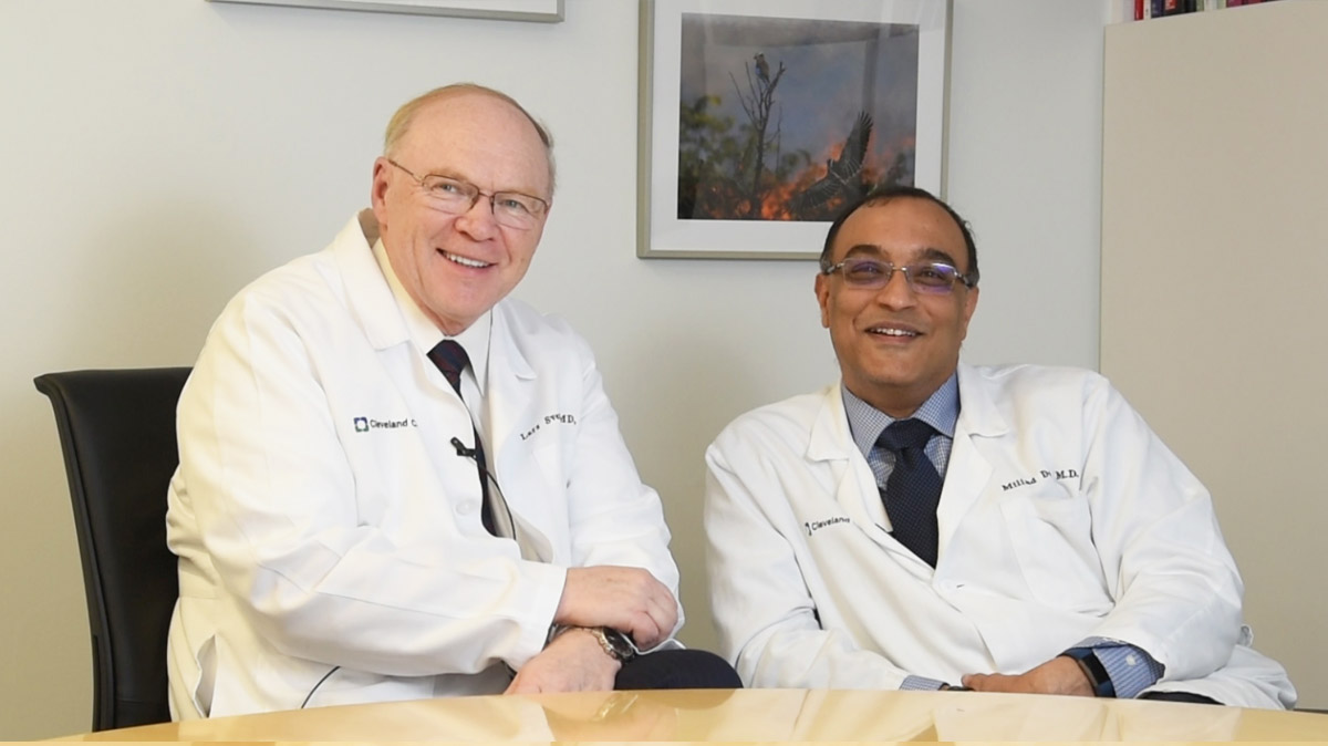
Dr. Lars Svensson, Chairman of the Sydell and Arnold Miller Heart and Vascular Institute discusses a recently published paper with Dr. Milind Desai, Medical Director of the Aorta Center, looking at a series of 491 Marfan Syndrome (MFS) patients who presented for both emergent and/or prophylactic aorta surgery. Dr. Svensson provides a review of the MFS guidelines regarding when to operate. Dr. Desai explains multi-modality imaging including echo, MRI and CT scan when determining timing for surgery. The findings of the study are reviewed as well as implications for future care of patients with MFS. Read the study in the Journal of the American College of Cardiology and learn more about Marfan syndrome at the Marfan Foundation.
Love your HeartSubscribe: Apple Podcasts | Buzzsprout | Spotify
Characteristics and Long Term Outcomes in Adults with Marfan Syndrome and Ascending Aorta Surgery
Podcast Transcript
Announcer: Welcome to love your heart, brought to you by Cleveland Clinics, Sydell and Arnold Miller, Family Heart and Vascular Institute. These podcasts will help you learn more about your heart, thoracic and vascular systems, ways to stay healthy, and information about diseases and treatment options. Enjoy.
Dr. Lars Svensson: Hello, I'm Lars Svensson, Chairman of the Heart and Vascular Institute, and also Director for the Marfan and Connective Tissue Disorders Clinic. That's something that's been my interest for many years and I'm here to work together with Milind Desai who's Head of our Umbrella and our Aorta Center. And we have been looking at patients who came here for a care of patients with Marfan Syndrome in particular. Now we obviously see a lot of patients with Loeys-Dietz and Ehlers-Danlos, and some of the more uncommon connective tissue disorders. But we particularly like to talk to you about the publication that is coming out now in the Journal of the American College of Cardiology, and something that Milind worked very hard and diligently to put together.
So let's just start off by talking a bit about the historical aspects of when to operate on patients with Marfans. So generally the guidelines have been above five centimeters unless somebody has a history of aortic dissection and in the HAACC guidelines, that I was involved in writing, we stuck to more or less five centimeters. We did talk a bit about cross-sectional areas of potential indication for surgery and then if patients had a history of dissection, operating earlier in patients. So Milind, what do you think about the whole issue of timing our surgery in Marfans and the notion of CAT scan versus Echo and various other criteria.
Dr. Milind Desai: Thank you very much Lars. So you know, I would like to address that in the bigger context of aortic disease in general, but also Marfan Syndrome. A lot of times in the past what we have relied upon is echocardiography to make measurements of aortic root and the ascending aorta. But as you know, e-hocardiography is a two-dimensional technique and often depending upon the ultrasound beam, if the beam is tangential, then your measurements can be off. You are not making accurate measurements and you are relying on diameters that may be erroneously obtained. So the world has generally shifted towards doing tomographic imaging, some sort of three-dimensional imaging, specifically contrast enhanced CT or MRI, when you are dealing with a such aortic pathology. Especially in patients with complex aortopathy histories like Marfan Syndrome, where the root pathology is very, very common.
And so you know, we routinely now... The world routinely now does a CT or MRIs because it gets rid of the tangentially, the ambiguity that comes with echocardiography. And here you can reconstruct a three dimensional data set in whatever plane you want and give precise measurements with less than a millimeter difference from study to study in terms of variability. So today I think if you have a patient with a aortic disease they deserve to have it come at least one tomographic scan to make sure we establish a baseline, and then decide the follow up.
Dr. Lars Svensson: So let me just follow up on what your comments were there about size and so on. Would you like to comment about CAT scans? Because remember CAT scans originally when we started doing them, we had historically been looking at chest X-rays and that's how we decided when to operate on patients. So we looked at a chest X-ray, and in those days it was usually say descending aorta six centimeters or bigger than chest X-ray, or abdominal, then we'd operate. So it was an outside diameter and echo. There's been talk about using the internal diameter or leading edge, leading edge, MRI. There's been some mixture there because MRI can be the external diameter versus MRA which is the internal diameter. You want to talk a bit more about those aspects?
Dr. Milind Desai: Yes. So I think it is important because you have to be comparing apples to apples and make sure you're not comparing apples to oranges in these situations. So by convention on Echocardiography, according to the ASC American Society of Echo Guidelines, you measure leading edge to leading edge during diastole. In CT, typically we measure at especially contrast enhanced CT, we measure the luminal diameter. So the numbers are not always going to be exactly similar. There are going to be discrepancies and the important thing, actually I forgot to mention is how crucial it is to do CAT scans with gating. Gating to the ECG triggers to Electrocardiography because you will have otherwise motion artifact of the root which will give you ambiguous reads measurements and that does not help anybody when you're making crucial decisions about precise measurements.
And the same concept applies in MRI. If you are doing an MRI angiogram, it needs to be gated, preferably with contrast. There are different techniques in MRI where you can obtain an MRI or angiogram without actually giving gadolinium contrast. But the important thing is gating is crucial and the measurement principals, you know luminal diameter, I think that's similar for all tomographic modalities. And often as you very well are aware, we are now moving away from diameter measurements. We are looking or trying to measure areas. We are actually rather than deriving areas from diameters, we are actually measuring areas because a lot of these aortas are eccentrically dilated. The roots are very eccentric and if you just use one diameter measurement then you may be off significantly. So these are some of the principles that you have to keep in mind when we are, you know, gating, calculate the area and understand if the root is symmetrical and circular then the diameter is okay. But if it is not, then you have to
be very careful about overestimating or underestimating that.
Dr. Lars Svensson: All right, so you touched a bit on area. Any thoughts about indexing when it concerns Marfan patients?
Dr. Milind Desai: I mean I would say indexing of the aortic root and the ascending aorta to height, in general for all aortic diseases, I think is absolutely crucial. Because you cannot have a the same diameter threshold for a six foot five athlete and a five foot two patient because the burden of a dilated aorta of the same dimension of aorta for a five foot two person may be very different compared to somebody who is six foot five. And the thresholds, in my opinion, especially based on diameter should be different. So you can level the playing field by indexing it to hide. And our work has shown that in multiple aortopathies and area to height ratio, the indexing area of the ascending aorta to height. And if the threshold is beyond 10 centimeters slash meter is associated with... could be used as a surrogate threshold for sending patients for preemptive surgery. And in our experience, longterm experience, if you do it that way, the survival is much better compared to relying on traditional thresholds.
Dr. Lars Svensson: Good. So let's talk a bit about the study now. So we started off looking at over a thousand patients here at the Cleveland Clinic who had a diagnosis of Marfans and we excluded patients under the age of 18 because we have a significant congenital population. In fact, that's how I started collecting the data 18 years ago as I took from one of our previous pediatricians who had an interest in Marfans, took those patients. It wasn't a big population, it was less than a hundred. And then we looked at all the patients who were operated, so over a thousand patients operated on. And then we went back and used a much stricter, more recent Ghent criteria to diagnose or used genetic identifiers of patients and mutations of patients who had Marfan Syndrome.
So we excluded Loeys-Dietz. We've just written a paper that's just come out about a week ago on Loeys-Dietz, 53 patients with Loeys-Dietz just to mention the Loeys-Dietz patients because there's relevance to Marfans. In the Loeys-Dietz patients that we operated on before they developed aortic dissection, the longterm survival was excellent. In the patients who we operated for Loeys-Dietz and they had aortic dissection, the longterm prognosis was not very good. So we had approximately 500 patients, just under 500 patients with Marfans and so strict criteria, diagnosis of Marfans and we divided again into the aortic dissection and the prophylactic surgery patients. You want to talk about that?
Dr. Milind Desai: Yeah, so thank you very much. And so it was 491 patients. And what as Dr Svensson mentioned, we wanted to have a very strict criteria because the criteria for Marfans have evolved and now there is the more newer revised Ghent criteria. So we retroactively applied the revised Ghent criteria to the larger data set of more than a thousand patients and the final number was about 491 patients and we divided them into two groups. Essentially the ones that came to us for prophylactic surgery on their dilated aortic roots, and the other ones who are transferred to us for an, eventual emergent surgery because they had a aortic dissection at an outside institution. So essentially, 299 patients in the prophylactic surgery group, 192 patients in the emergent surgery group.
And the important thing, as I said, one was the revised Ghent criteria was strictly applied. Number two, 93% of these patients had a CAT scan or a cardiac MRI at our center. So gated high quality CAT scan or a high quality MRI. And the other 7% had, because it was in a dire emergency, we relied on the outside CT scan reads. And pretty much everybody had at least some form of a baseline echocardiogram performed before they went to surgery. So big picture was all these patients were operated. Importantly in the prophylactic surgery group, about 53% patients underwent valve sparing root replacement. So a very advanced slick surgery was performed with the intent, because a lot of these patients are younger we want to preserve their valves, so we did a valve sparing root surgery. And obviously in the emergent surgery group, majority of them underwent composite valve graft or some sort of salivate situation.
We followed these patients and there were 14 deaths. Very small proportion or the survival in hospital survival was more than more than 98% and we followed these patients for a median of eight years. And what we have shown is that patients, who on multivariate analysis, patients who underwent a prophylactic surgery, their outcomes were significantly better compared to the patients who were operated emergently. That's not a surprise. The other thing we also found was, especially in the prophylactic surgery group, the folks who underwent valve sparing root replacement, their longer term outcomes were better than the composite valve surgery. The other crucial point of our study was we specifically then looked at the 192 patients who had emergent surgery and what we found is on the day of their presentation, on the day of their surgery, 56% patients did not meet the current guideline recommended size thresholds. They were under the guideline recommended size threshold for surgery. Meaning if they had arrived at o
ur institution the day before their dissection and we look at the CAT scans or their MRIs, they would not have met threshold for guidelines with relation to their surgery.
So that begets the question, or that leads us to the following few observations. One, if you are a patient with Marfan Syndrome, you should be plugged into an experience center, not just an experience medical center, but an experience center that can provide the full spectrum including high quality medical care, high quality imaging, high quality surgery, and high quality genetic care. The other thing is, especially this study raises the question, should we be looking at lower size thresholds for patients with Marfans for prophylactic surgery, particularly the way it relates to in an experience center. So I think these two were two big important points that I got from the study.
Dr. Lars Svensson: Yeah, so I think that the important message here was that in the patients who had emergency surgery, the results were actually very good. We had a 3% risk of death. Whereas overall for the country, for acute dissections and mortality rounds in that I read database 25% more, one 17%, our own mortality rate here for acute dissections, for everybody at the Cleveland clinic, usually runs about 6%. But in this Marfan group, obviously younger, it was 3%. In the patients who had the prophylactic operations, and we do a modification on the day of the operation. The operation that I think is much better, particularly in Marfans because you're less likely to have tears in the mitral valve base, in the aortic root, were that we use pledgers. We reduce the size of the aortic annulus by two the size it should be for a person of equivalent height.
We've actually now done over 870 reimplantations, not all of them obviously in the Marfan population with, we've got a paper that we working on now that looks like the durability is going to be about 97% freedom from reoperation at 10 years. And we're going to actually look at our Marfan patients versus the patients who don't have Marfans and do a study on that because the big concern has always been, in patients with Marfans, we know when we used to take the leaflets out, they showed what we call myxomatous degeneration. And that's why there was a reluctance by Vincent Gott who was at Johns Hopkins about doing reimplantations in the Marfan population.
Now getting back to the issue of timing of surgery, we obviously have in a sensor numerate of the number of patients we saw with an an aorta that was less than the recommended guidelines for operating on. We don't have the denominator. In other words, how many patients with Marfans dissected with those sizes.
However, when I was in Boston, I did have a population of Marfan patients I followed and what was interesting was that 15% of those patients dissected at a size less than five centimeters. And that's what resulted in me coming up with this sort of a cross sectional area to height ratio. In other words, if the cross sectional area in square centimeters is divided by the patient's height, and if that ratio is more than 10, we recommend surgery. In Vincent Gott's experience, same thing. In the Johns Hopkins study from many years ago, 15% of patients dissected at less than five centimeters. So here we have a study now showing that a lot of patients are dissecting at a size less than five centimeters. Obviously again, we don't know what the total population is, but we perhaps should give more thought to that.
And particularly look at the patients who have a history of aortic dissection. I think it's very important as a patient that has a history of aortic dissection, we operate earlier. And then the longterm results are already very good for both populations. When I was in Houston, I wrote a paper on Marfans and essentially a lot of them had reoperations and by five years, only 75% were still alive.
In this study, we've now shown that at eight years, 98 sorry, at eight years, 85% of patients were alive who had aortic dissection. In the patients that had prophylactic surgery, 92% was still alive. So that gives you some idea of the issues. Now, one of the things that we're obviously still concerned about is in patients where we do a prophylactic reimplantation operation, we know the results are excellent, survival's excellent. But down the road there are some patients who develop aortic dissection and we looked at our total population of reimplantation and said, what's the risk of later developing aortic dissection, usually the descending aorta. And we found that it was 1.4% and a risk factor for dissection after reimplantation was a connective tissue disorder, maybe a bit higher in patients with Loeys-Dietz. So all in all I think this shows excellent results and that the reimplantation operation is very good. Obviously you're saving a patient's valve for them. They don't have to go onto being on Coumadin for a composite valve graft. Obviously most of these patients are young patients. Any final comments, Milind?
Dr. Milind Desai: So I think this again points to teamwork experience and the value that a high volume experience center that works seamlessly brings to the table. I mean, and as I alluded to earlier, it's not just that there's a good cardiologist, there is a team of people that are working in synchrony. And even for the patients who are unfortunate to have a dissection and then fly here, our acute aortic team works seamlessly to push the envelope. The boundaries of what we can provide to these patients. Not just Marfans, but anybody that presents with an aortic dissection. And some of the amazing work we are doing, complex surgeries in really sick patients with excellent results. So I think this, to me the big message of this is teamwork is crucial for the success of any program.
And the other thing the rebel in me always likes to challenge the status quo and pushing the envelope in terms of you know where are these guidelines coming from? What are these recommendations coming from? Should we be evolving in terms of these guidelines as technology improves, as our understanding occurs. So I think the stat, I like to challenge the status quo, and this is one other example where we seem, I think we should be, we should be relooking at some of the size thresholds.
Dr. Lars Svensson: Well good. Thanks very much Milind. I think we are going to see evolution of the guidelines. I've talked about updating them. We may do a smaller document on updating the guidelines and that includes obviously connective tissue disorder management. There's been a lot of new information on the genetic side that is very useful. There are a couple of prospective randomized trials including one we did here on brain protection and there's no doubt about the whole field is going to evolve. It's not only genetics, it's proteonomics, it's metabolics. We have a lot of research going on to the genetic side here at the Cleveland Clinic and also some of the metabolic side of things. And ultimately hopefully we can see a patient with Marfans and say, based on your MRI, you show these kind of proteins, probably some proteoglycans in your aortic wall. You've got a family history of this, this is your gene. And so we believe we can keep on watching you or we need to operate on you. That's the future that we look forward to.
Dr. Milind Desai: And I think the other, to dovetail into that. The other important aspect is artificial intelligence. So there is going to come a time when we can potentially predict, based on artificial intelligence and serial scans, who is at a higher risk, what have you, on top of all the other omix that doctor Svensson talked about. So I think the future is exciting.
Dr. Lars Svensson: Yeah. We'll be number crunching and putting it all into a machine and machine learning hopefully will spit out answers for your future. Thank you very much for listing and being part of this.
Dr. Milind Desai: Thank you so much.
Announcer: Thank you for listening. We hope you enjoyed the podcast. We welcome your comments and feedback. Please contact us at heartatccf.org. Like what you heard? Please subscribe and share the link on iTunes.

Love Your Heart
A Cleveland Clinic podcast to help you learn more about heart and vascular disease and conditions affecting your chest. We explore prevention, diagnostic tests, medical and surgical treatments, new innovations and more.


