A Look at Echocardiography

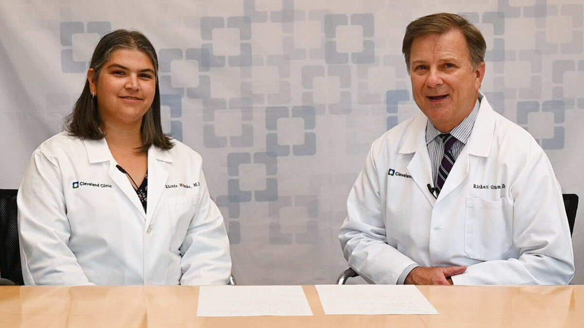
Echocardiography can be a useful diagnostic tool in caring for cardiac patients. Dr. Richard Grimm and Dr. Rhonda Miyasaka speak about various types of tests using echocardiography. Learn indications, benefits, and challenges of these imaging tests.
Subscribe: Apple Podcasts | Buzzsprout | Spotify
A Look at Echocardiography
Podcast Transcript
Announcer:
Welcome to Love Your Heart, brought to you by Cleveland Clinic's Sydell and Arnold Miller Family Heart, Vascular and Thoracic Institute. These podcasts will help you learn more about your heart, thoracic and vascular systems, ways to stay healthy, and information about diseases and treatment options. Enjoy.
Richard Grimm, DO:
Good afternoon, my name is Rick Grimm. I'm a cardiologist and Director of the Echocardiography Laboratory here at Cleveland Clinic, and I'm here with one of my partners, Rhonda Miyasaka, who is Director of our Interventional Echocardiography team here at Cleveland Clinic, and we're here to tell you all about the world of echocardiography, which I believe is one of the most important diagnostic procedures in our cardiovascular realm of techniques and procedures to date.
Richard Grimm, DO:
Echocardiography, for those of you who may not be aware, is a noninvasive test in which we use echo Doppler technology to actually assess heart size, function, valvular disease, valvular regurgitation, narrowings, as well as the hemodynamics and pressures that are generated by the heart to determine and to evaluate for any pathology. This is something that we do in our laboratory quite often, it's probably the most common diagnostic procedure performed in cardiology today. Other such cardiac tests include electrocardiograms, that look at the heart rate and rhythm, as well as on the other end of the spectrum, cardiac catheterization or invasive cardiology techniques that actually look and define and examine the arteries that supply the heart muscle itself.
Richard Grimm, DO:
An echocardiogram is a test, again, that's completely noninvasive. We literally generate images from the chest wall with an ultrasound probe, and we're able to evaluate and diagnose many pathologies in patients that are having one ordered as a result of an evaluation, an initial examination or evaluation, either by their general family practitioner, internist, or a cardiologist as well. I'm going to ask Rhonda to talk a little bit more about the evaluation in general, as well as the examination itself.
Rhonda Miyasaka, MD:
Sure. Thanks, Rick. Thanks for having me here today, this is one of my favorite topics to talk about, is actually echocardiography, or ultrasound of the heart. And one of the things that we'd like to make sure that we talk about are some of the common questions that we get from patients. So when your doctor orders an echo, why did they order the echo? Why do you need an echo? Some very common reasons why your doctor may have ordered an echo would be maybe you reported a symptom that you were having, perhaps some chest discomfort or shortness of breath, and your doctor wants to make sure that everything looks okay with your heart. Perhaps they noticed an abnormal sound when they listened to your chest, like a murmur, an echocardiogram is very good at trying to understand the reason why you may have a murmur. Or maybe they ordered it because they found another test that looked abnormal, like a blood test, or a chest x-ray, or an EKG. So it's a very helpful tool to be able to understand how your heart's doing.
Rhonda Miyasaka, MD:
From an echo, we can get a lot of really great information. We can understand your heart size, we can understand your heart function, we get a detailed look at all of the valves, we can make sure they're opening okay, they're closing okay. We can do measurements. And so it's a really, really valuable tool that we have in cardiology. Rick, maybe to kind of bring things back over to you, what should our patients expect? You've ordered an echo in clinic, and they come to the check in desk, what is an echo like for them? Who does it? How long does it take? Are there any risks that they have to worry about?
Richard Grimm, DO:
Right. Well, the beauty of this technology is that, again, it is noninvasive. There are virtually, there are no risks to the procedure itself. It literally is a test where an ultrasound probe, which is about the size of a microphone, is actually applied to the chest wall with a little bit of gel, and images are acquired by a cardiac sonographer. This is a technician that is specifically trained and educated on the acquisition of echocardiographic or ultrasound images from the chest wall. These technicians are extremely highly knowledgeable, highly skilled, and are very, very, very well trained. And especially in a place like the Cleveland Clinic, in our laboratory, where we see a tremendous amount of pathology, and certainly a large number of patients, and these sonographers as a result of performing large volumes of studies are outstanding at what they do in terms of acquiring images.
Richard Grimm, DO:
Once these images are acquired, and usually this test takes approximately 45 minutes to conduct, sometimes a little less if there are no abnormalities whatsoever, sometimes a little longer if we find particular areas of interest or concern. And certainly this technique is very much impacted by a variety of things, such as a patient's size, for example, ultrasound is a wave form that has difficulty penetrating through large masses, so large body sizes tend to be challenging for us, and that may take a little longer in terms of that interrogation.
Richard Grimm, DO:
But in any case, and even though this is a noninvasive procedure, that seems to signal to many of the non-inclined that it's not a very high expertise examination. And counter to that, this technique is actually extremely useful for us and provides us a tremendous amount of information and data whenever it's performed. But it can be adversely impacted, again, by technical challenges in patients, again, such as body size and actually lung disease, for example, that can be a challenge for us in patients that have significant end stage lung disease, as an example.
Richard Grimm, DO:
Again, these tests can be applied in different environments as well. It's extremely portable, we can take it anywhere. We can acquire images in literally seconds, and in real time, and as a result, it can be extremely important for us in making diagnoses that may be time sensitive, and very accurately and effectively. So it's a seemingly simple technology that has tremendous ramifications and capabilities in terms of being extremely powerful in its ability to make the diagnosis of significant disease.
Rhonda Miyasaka, MD:
Maybe something else to comment on as well, patients are always worried, is there anything I have to worry about during the test or the procedure? Am I going to need an IV? Do I need blood work? Maybe we can help just alleviate some of those concerns as well. Most echoes, we don't have to do any particular blood work for screening, there really aren't any major risks to it. Sometimes we do end up needing to use an IV, and that is for some more specialized types of imaging. Maybe you could talk about the situations where we do things like a bubble study and what that means, if a patient says we need to do one of those, or echo contrast, and what that means because the word contrast sometimes has some connotations for people.
Richard Grimm, DO:
Right. Well, just as in other radiologic procedures where a contrast agent may be utilized to enhance certain features of that test, the same is true with echocardiography, although the contrast that is utilized is very different from the iodine directed dye that's used, for example, in radiologic circles with CAT scans, for an example. Our contrast agents are really comprised of bubbles, small microscopic bubbles that actually can enhance the ultrasound image. Additionally, we can use and inject these, what we refer to as agitated saline, which is very harmless, and can be injected, but also enhances the ultrasound signals, and can, in fact, allow us to detect and identify shunts in the heart when communications exist between the left and the right side of the heart that shouldn't under normal circumstances.
Richard Grimm, DO:
So we do have other techniques and it can be used in other scenarios as well, such as stress testing. We utilize it to evaluate the heart function before and after stress testing. And then we have another capability that is particularly advanced, and that is our transesophageal echocardiographic exam, where we have an ultrasound probe on the end of an endoscope that we can actually insert into the food pipe, just like an endoscopy, and take exquisite images of the heart, literally because the probe is sitting right in the esophagus, right adjacent to the heart, behind the heart. And maybe Rhonda can tell us a little more about that capability which is really just booming and a tremendous capability for us as well.
Rhonda Miyasaka, MD:
Sure, absolutely. So what Rick was talking about is a test that we call a transesophageal echo, or T-E-E. And it's the same type of technology in that it uses ultrasound to get really nice pictures of the heart. But the benefit is that this test is actually done as a procedure, meaning that we give some sedation and some numbing to the throat to make sure that our patients are nice and comfortable, and then we just very simply advance a small ultrasound probe down the throat so that we can take really close up pictures of the heart. The advantage of a transesophageal echo is that our image quality is absolutely pristine. And so, sometimes, once we get the regular type of transthoracic echo, or the type we've already talked about, where we have the wand that we wave on the outside of the chest, a transesophageal echo can give us much finer details, particularly if we need to understand issues or problems with valves. That's one of the main reasons why we would do transesophageal echo is to get really nice pictures of your valves.
Rhonda Miyasaka, MD:
So your heart had has four different valves, and on T-E-E we can get very crisp images, we can understand what the leaflets look like, we can understand how will they open, how will they close. We can see if there are areas of leakiness, or areas of narrowing. And the technology these days is absolutely amazing because we now have the ability to, in addition to getting our regular echo views, we can actually generate three dimensional images. So three dimensional images of the heart, three dimensional images of the valves, and we can rotate the valves and make them look almost like a surgeon would be looking them as if they were able to look right into your chest. And so it gives us a really great understanding of what the valves look like, and it helps us understand if you might need a treatment for those valves, whether that be a medication, or a procedure, or surgery.
Rhonda Miyasaka, MD:
Another reason that we do a transesophageal echo, for those of our patients that have heart rhythm abnormalities, like atrial fibrillation or atrial flutter, sometimes we'll do a transesophageal echo to look at the heart closely and make sure there are no blood clots before a procedure called a cardioversion, or a shock treatment. And so it's a test, again, that we do very commonly here, but it gives us really nice information and it's very complementary to the regular echo that you probably had beforehand.
Rhonda Miyasaka, MD:
How about maybe some questions that patients ask me all the time? We see a lot of patients that have had echoes in the past, they've had an echo close to home, and now they're coming to see us for the first time, and sometimes we say, "Hey, I know you already had echo, but we want to do another one." Why might we sometimes want to do another echo? And along those lines, as the head of our echo lab, what are some things that you're proud of that we're able to do here, and offer here, as part of the echo experience?
Richard Grimm, DO:
Well, I'll take the second question first, and thank you for that lead in. But I think, certainly here at Cleveland Clinic, as I mentioned, we do a tremendous volume, and it's probably a larger volume than most places anywhere. And as a result of that, again, not only our sonographers but our professional staff have tremendous experience and expertise in evaluating these cases that often are very, very complex cases. One of the reasons why a patient may need a repeated exam is that if an exam has been performed elsewhere, there's a possibility that they may have certainly had someone with lesser experience or expertise performing an exam. It may not have been on the highest quality equipment, which makes a tremendous difference in terms of image quality, and oftentimes the examinations may not be as extensive and in depth.
Richard Grimm, DO:
And there are lot of capabilities, as I mentioned earlier, that we can do with echocardiography and Doppler echocardiography, where we can make measurements and actually interrogate, noninvasively, pressures within the heart, and that can give us a tremendous amount of information in and of itself. And we can really perform directed examinations, directed to the problem at hand, and that is not always conducted in a general ultrasound exam. I think in most, I'd like to say, and it is very, very true, it's a very stark difference, but in most places that perform echocardiography, I'd say a vast majority, maybe 60 to 80% of their examinations are essentially normal exams. Here at the Cleveland Clinic in our laboratory, it's just the opposite, 80% of our examinations are abnormal and have significant pathology. So it's all around us, we're very commonly seeing these, very well aware of the potential of a lot of these. Again, the immediate feedback that we have enable us to act fairly quickly on issues that may require more urgent evaluation, and we can then refer on to other areas, as necessary. So again, an incredibly valuable technique.
Richard Grimm, DO:
The other area that I think many may not be as familiar with, is the fact that, as I mentioned, it can be utilized in other circumstances more acutely. Again, in the hospital environment, you'll see that it's utilized quite frequently on the wards, in the intensive care units, and to guide other procedures. That's something both in the operating room that is commonly utilized, as well as in the outpatient department, as well as in the cath lab, for example, to guide procedures that can be done less invasively. And that's something that Rhonda has been particularly expert in helping our team with here at Cleveland Clinic as well. But all in all, a remarkable technology, the rest of the world has become familiar with it, so all areas outside of cardiology are now more routinely utilizing this technique, and it's a wonderful technique to have available in our armamentarium.
Announcer:
Thank you for listening. We hope you enjoyed the podcast. We welcome your comments and feedback. Please contact us at heart@ccf.org. Like what you heard? Subscribe wherever you get your podcasts, or listen at clevelandclinic.org/loveyourheartpodcast.

Love Your Heart
A Cleveland Clinic podcast to help you learn more about heart and vascular disease and conditions affecting your chest. We explore prevention, diagnostic tests, medical and surgical treatments, new innovations and more.


