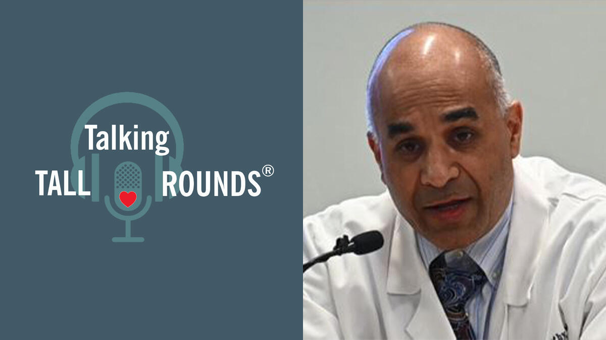
Talking Tall Rounds®: Incidentally Discovered Lung Nodules during Cardiac Testing

Dr. Sudish Murthy discusses the management of incidentally discovered lung nodules during cardiac testing.
Enjoy the full Tall Rounds® & earn free CME
- Case Presentation: Andrew Feczko, MD
- Incidence, Institutional Strategy for Incidental Nodules: Peter Mazzone, MD
- Reading the Lung Part of a Cardiac Test - Radiologist’s Perspective: Michael Bolen, MD
- Surgical Management of Indeterminate Lung Nodule: Usman Ahmad, MD
- Pulmonary Surgery in Cardiac High Risk Patient: Daniel Raymond, MD
- Non-Surgical Treatment of Lung Cancer - Radiation Oncology Perspective: Gregory Videtic, MD
- Timing Pulmonary Surgery after TAVR and Coronary Stenting - New Data: Amar Krishnaswamy, MD
Subscribe: Apple Podcasts | Buzzsprout | Spotify
Talking Tall Rounds®: Incidentally Discovered Lung Nodules during Cardiac Testing
Podcast Transcript
Announcer:
Welcome to the Talking Tall Rounds series brought to you by the Sydell and Arnold Miller Family Heart, Vascular & Thoracic Institute at Cleveland Clinic.
Sudish Murthy, MD, PhD:
Welcome to Heart, Vascular & Thoracic Institute Tall Rounds. This session is dedicated to the management of incidentally discovered lung nodules on cardiac testing. This is a surprisingly common problem for us and directly seems to impact the H and V faculty and indirectly saddles the T faculty. So today we're going to present a case and share some of our thoughts with you.
Sudish Murthy, MD, PhD:
Our first introductory talk will be given by Andrew Feczko. He's our chief resident in general thoracic surgery who will start with our case presentation.
Andrew Feczko, MD:
As Dr. Murthy mentioned, my name is Andrew Feczko. I'm one of the thoracic surgery residents. I'll be presenting a case that illustrates some of the considerations involved in managing these incidentally identified pulmonary nodules during cardiac testing. So this patient was an 81 year old former smoker who initially presented to the Cleveland Clinic for management of severe aortic stenosis that was identified during workup for a syncopal episode. He was initially treated with a balloon valvuloplasty and then referred for consideration of valve replacement. However, during workup preoperative CTA demonstrated a 3.9x3.3 centimeter left upper lobe mass, which was ultimately identified as a clinical T2aN0 adenocarcinoma. Through cardiac surgery evaluation he was deemed to be an acceptable risk for open heart surgery. However, a TAVR was favored in this case to facilitate expeditious treatment of his incidentally identified lung cancer. On the 14th of June in 2017, he underwent a transfemoral placement of a 29 millimeter Edwards SAPIEN aortic valve prosthesis.
Andrew Feczko, MD:
And then in July of that year, about three weeks later, underwent a robotic-assisted left upper lobectomy and mediastinal lymph node dissection. He recovered very well from this was discharged on postoperative day number two with a pathologic T2aN0 adenocarcinoma as his final pathology. He didn't undergo any adjuvant therapy at that time and has been continued on surveillance, both for his valve replacement as well as for his lung cancer. He has been identified to have bilateral pulmonary nodules, which have occurred in the last year, and is currently undergoing consideration for systemic therapy for those.
Andrew Feczko, MD:
So as a couple discussion points, this case demonstrates the challenges that are created by synchronous identification of multiple thoracic pathologies and the importance of a multidisciplinary approach and the application of advanced technologies to adequately address all these issues in a patient-centered fashion. This scenario will continue to be identified as we rely on axial imaging for screening and diagnosis and surveillance of these pathologies. Disease-specific surveillance obviously remains an important part of the post operative care of these patients and, again, requires a multidisciplinary approach.
Usman Ahmad, MD:
Thank you very much. Good morning.
Usman Ahmad, MD:
Thank you, Dr. Murthy and the Tall Rounds team for organizing this very important debate and elaboration on what we do for these patients. Because if you think about it, most patients with heart or lung disease who walk into a hospital have a significant overlap of risk factors for the other disease. So when taking care of a heart or a lung problem, it's really important for the providers to pay attention to the other organ, make sure we don't skip or miss the disease for which the patient has significant risk factors for or they may.
Usman Ahmad, MD:
Before I move forward, I want to acknowledge and thank the contribution of Naomi Bolen. That was fantastic work there. Anyway, so surgery maintained an important role in management of lung nodules, particularly since we're worried about presence of malignancy and lung cancer, given the risk factors in our patient population.
Usman Ahmad, MD:
If you look at the trajectory of survival in lung cancer patients, there are two things that are quite obvious. This is data from the IASLC TNM staging work, looking at international data on the right side of the screen and US data on the left side of the screen. The two things that stand out are, 1) We've really not made a whole lot of progress in improving survival in all-comers for lung cancer. So if you take all lung cancers together at five years, only a fifth of those patients or 20% will be alive, which is quite poor and dismal compared to other solid organ tumors. The other important fact here is that if we can catch the tumors early and treat them early with either radiation or surgery, their survival actually is quite good at reaching up to 70-80% at five years, which is quite remarkable.
Usman Ahmad, MD:
And some here would even go out to argue and propose that in early stage patients and stage 1a patients, they're more likely to die of "natural causes" than lung cancer if we can treat, manage and surveil their lung cancer appropriately. So the emphasis here then becomes on early detection and early treatment of lung cancer. This really received more credence and a lot of steam after the National Lung Cancer Screening Trial came out in 2011 in the New England Journal. And you see the now famous graphs here on the left side of the screen, which showed that with early detection in high risk patients, lung cancer, mortality was decreased by 20%, which is really unheard of, compared to breast cancer, colon cancer, or any other solid organ tumors.
Usman Ahmad, MD:
And since more CTs were done, investigators in other institutions and us, as Dr. Mazzone had pointed out a few minutes ago, figured out strategies to classify these nodules into high-risk and low-risk categories so they can be intervened upon and followed appropriately, which led to this development of Lung-RADS protocol, similar to the breast BI-RADS protocol, which differentiates the nodule into high-risk un-intervenable nodules and those that can be followed.
Usman Ahmad, MD:
So when we see these indeterminate pulmonary nodules as Dr. Mazzone and Dr. Bolin pointed out, there's a whole array of diagnoses that can be associated with them and really have to think about and work through the algorithms carefully to identify patients appropriately for appropriate treatment strategies or surveillance. So should we continue to monitor these indeterminate nodules? And if so, at what intervals should they be biopsied? And if so, should these biopsies be percutaneous, image-guided, or bronchoscopic? Should they have surgery? And then, what surgery? Or should they have radiation, with or without a biopsy first? So these are many different question that are sometimes difficult or challenging to address and answer, and really require input from all members of the treating physician team and provider team, in a multidisciplinary fashion. And that's really the key in managing these indeterminate pulmonary nodules to put all the heads together from all these various specialties to try to come up with an optimum plan for treatment.
Usman Ahmad, MD:
This is an example of what monthly disciplinary selection and management does. So this is data from the Lahey Clinic, recently published, that looked at a protocol very similar to what Dr. Mazzone had described and has established here. And I want to thank him for his leadership in building up this program at the Cleveland Clinic. In their program, they identified 354 high-risk nodules and, after appropriate pulmonary and multidisciplinary consultation, they submitted a select portion of them to surgery and, with appropriate selection, less than 1% of the nodules had surgery for benign diseases and most of the nodules that were resected ended up being lung cancers, which is a very important piece of data here. And it's important to highlight that with appropriate selection we don't necessarily need to do unnecessary surgeries on these patients. And that is really the key in establishing such protocols in any institution. And we're happy to report that we've done that here at the Clinic.
Usman Ahmad, MD:
So when we talk about surgeries for lung cancers, the vast majority of the surgeries done here are some sort of minimally invasive surgery, either thoracoscopic or robotic, which means that the patients have low morbidity or quick recovery with equal oncologic legitimacy of the operation as we've shown in our data multiple times.
Usman Ahmad, MD:
So I'll just discuss with you briefly some of the innovations that we've incorporated in achieving these high rates of minimally invasive surgery. So one of the two techniques that I'll share with you here today is this specialized needle localization or microcoil localization that is performed by Dr. Bolen's group and some of the other interventional radiologists. So here's an example of a patient with a peripheral, small nodule that would be impossible for us to feel or see thoracoscopically or robotically. So in these situations, we ask our radiology colleagues to access the nodule on the day of surgery. Here the patient is upside down and you can see this needle being put into the nodule and through that needle they deploy a spring type microcoil, part of which is in the nodule and part of it is at the surface of the lung, allowing me and the other surgeons to see the nodule during surgery.
Usman Ahmad, MD:
And this is what it looks like during surgery. So now the camera is in place. You can see that the lung is deflated. The ribs are on the side here. And in the lower lobe, the lesion that you saw on that CT scan has been marked by that microcoil. And there you see the coil, which is now being... There's a stitch being placed through the nodule under the coil to lift it up to resect. So this is a robotic procedure--you can see the robotic instruments--allowing us to do this sort of thing with quite a bit of dexterity.
Usman Ahmad, MD:
And after the nodule is identified, lifted up and we can resect it using our special resectional devices. This is an endo-stapler, which staples the edge of the lung that needs to be resected and also transects it. And we can use several of them to finish the procedure. Here this nodule is separated with a nice rim of lung around it. Usually we would test this nodule during surgery, and if it is a lung cancer, we would proceed with a formal anatomic lung cancer resection in the same setting. If it ends up being a benign nodule, then this is a very low morbidity procedure that the patient can essentially go home from the following day and recover really without any problems.
Usman Ahmad, MD:
The other innovative technique that we are part of a clinical trial for now, moving on, we were actually part of the Phase 1 trial now in the Phase 2 setting, is a tumor marker in fluoroscope conjugate, OTL-38, which is a folate analog and for this particular study is bound to a fluoro-4, which can be detected with near infrared imaging intraoperatively, and allows us to find these nodules there towards the surface of the lung during surgery.
Usman Ahmad, MD:
So the patients receive this folate-receptor conjugate preoperatively, a few hours before surgery. And then during surgery, we can find the nodule with fluoroscopy and resect it. So here is an example of what this looks like. Again, this is minimally invasive procedure. The lung is deflated. You can see some nonspecific feedback from the chest wall and from other portions of the lung, but as we focus on the nodule under question here, you'll start to see that it becomes obvious quite clearly. So here's the nodule under question, you can actually see the puckering in the visceral pleura here, and you can see quite a bit of uptake of the dye and the fluorescent feedback from it. So this is how we've actually detected nodules that were not detected on patients' preoperative CT scans and resected them during surgery. Now we're looking at other parts of the lung and don't really significant see any significant uptake in other areas.
Usman Ahmad, MD:
So these are some of the innovations that we've adopted to improve the role of surgery, the conduct of surgery, and decrease the morbidity of surgery. And we're very excited to have these innovative strategies in our portfolio and in our armamentarium. And we'll continue to push forward with these and we'll share with you our findings once these trials are finalized. Thank you.
Announcer:
Thank you for listening. We hope you enjoyed the podcast. Like what you heard? Visit Tall Rounds online at clevelandclinic.org/tallrounds and subscribe for free access to more education on the go.

Cardiac Consult
A Cleveland Clinic podcast exploring heart, vascular and thoracic topics of interest to healthcare providers: medical and surgical treatments, diagnostic testing, medical conditions, and research, technology and practice issues.


