Talking Tall Rounds®: Adult Congenital Heart Disease & Pulmonary Hypertension Combined Clinic

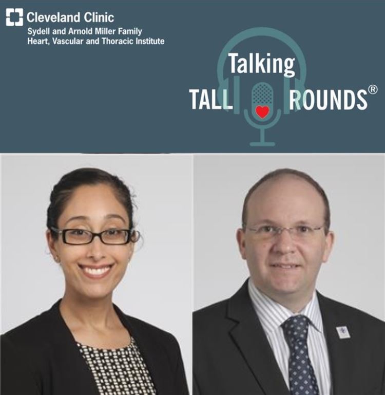
Tamanna Singh, MD and Adriano Tonelli, MD discuss non-invasive and invasive cardiopulmonary exercise testing.
Enjoy the full Tall Rounds® and earn free CME
- Introduction by Moderator: Gosta Pettersson, MD, PhD
- Case Presentations: Nicole Pristera, MD
- Evaluating Suspected Pulmonary Hypertension: Neal Chaisson, MD
- Cardiopulmonary Exercise Testing - Noninvasive: Tamanna Singh, MD/ Invasive: Adriano Tonelli, MD
- Heart Failure Management Insights: Sanjeeb Bhattacharya, MD
- Interventions for ACHD-PH: Joanna Ghobrial, MD
- Surgical Approaches: Michael Tong, MD
Subscribe: Apple Podcasts | Buzzsprout | Spotify
Talking Tall Rounds®: Adult Congenital Heart Disease & Pulmonary Hypertension Combined Clinic
Podcast Transcript
Announcer:
Welcome to the Talking Tall Rounds series brought to you by the Sydell and Arnold Miller Family Heart, Vascular and Thoracic Institute at Cleveland Clinic.
Tamanna Singh, MD:
Thanks everybody for having me this morning. So I'll talk, cardiopulmonary exercise testing is a huge topic, so I'll try to do what I can to just introduce you to this type of modality from a noninvasive perspective and how it's applicable to the congenital heart disease population and CHD-PH population. In addition to that, we'll also talk a little bit about just the mechanisms of exercise tolerance.
Tamanna Singh, MD:
So, the basics about cardiopulmonary exercise testing it's quite useful with respect to assessing the amount of inhaled oxygen and expired carbon dioxide, the gas exchange during stress, and we can actually assess three different types of systems: the cardiovascular, pulmonary, as well as neuromuscular systems. And it's very useful for actually being able to objectify exercise capacity. Other types of stress tests are a little more subjective in my opinion, so I think that these can be more reliant and actually are quite accurate when we're trying to compare the estimated peak oxygen consumption to that which is measured.
Tamanna Singh, MD:
And then it's also very useful, not just for risk stratification, but also for prognostication when we're talking about our CHD population, as well as heart failure and clinical decline in that regard. So, when we do cardiopulmonary exercise testing, it's really important to tailor the modality and exercise intensity to not just the individual's athletic ability. These are congenital patients, but they come with a variety with respect to what their capabilities are, and yes, perception can play a part in that, and their protocol should be something that's within an eight to 12-minute period. And this is typically what we look at when we're evaluating the data, and these are just four plots of a nine-plot panel that we typically get in our report. And I just show this to you if you ever want to familiarize yourself with the results.
Tamanna Singh, MD:
We look at peak oxygen consumption, the top left, we look at minute ventilation in the top right, and we take a look at the responses of heart rate and oxygen pulse which can sometimes be equated to surrogate for stroke volume in these patients, and then the overall just gas exchange response, comparing oxygen uptake and carbon dioxide elimination. So in our congenital heart disease population, exercise intolerance is really affected by a multitude of mechanisms, both being non cardiac, as well as cardiac. And this was just a nice pictorial describing that going from outflow obstruction and valve disease, all the way to arrhythmogenic etiologies and medications, pressure-volume overload, and then eventually going into the pitfalls of ventricular dysfunction, pulmonary vascular disease, and also incorporating some musculoskeletal abnormalities. We can see scoliosis for instance, sometimes in this population and then common things such as anemia.
Tamanna Singh, MD:
And I think, again, it's very important to emphasize that patients themselves may underestimate their exercise tolerance because they're born with a specific expectation, and that's not necessarily the same as our non-congenital population. And because of that, there's often a discrepancy between our subjective NYHA classification and their actual true exercise capacity measured by cardiopulmonary exercise testing.
Tamanna Singh, MD:
So in our congenital heart disease population, these are some primary indications for CPET in this population, predominantly to assess physical capacity, which is a main goal. And then to also evaluate causes of exercise and do symptoms, respect stratification and prognostication, and then to evaluate the need for medical therapy and the success of medical therapy, the potential for success with surgical or percutaneous procedures and post-repair, as well as the need for a heart transplant or heart and lung transplant. It can also be very useful when we're trying to prescribe a quote-unquote exercise prescription or provide some recommendations that they can take to cardiac rehab. And it's also very useful to reassess their growth after they do complete cardiac rehab either before or after therapeutic or surgical modality.
Tamanna Singh, MD:
The greatest value like I alluded to is really the ability to objectively determine their maximal effort. And we use a specific parameter, the respiratory exchange ratio, to be able to define whether or not they're able to extend their exercise capacity into the anaerobic metabolic state. So the next couple of slides are a little bit busy in the sense of having a great deal of text to them. But again, I wanted to introduce you to the parameters, and the definitions that we use in case you do look at these reports and are trying to decipher them for yourselves and understand how they may be relevant to your patients and how they can be useful to determine next steps in management.
Tamanna Singh, MD:
So peak VO2 is our oxygen consumption. And as you can imagine, it's quite lower in our congenital population than those who are healthy, and it can be very predictive of all-cause mortality hospitalizations, as well as adverse outcomes. Heart rates in these populations tend to be blunted due to an element of autonomic dysfunction. And these patients typically reach about 30 to 60% of their max predicted heart rate. And then they also come with a slew of arrhythmias that can be very common either before or after repair.
Tamanna Singh, MD:
Heart rate reserve, which we define as the difference between peak heart rate and heart rate at specific recovery times can also be compromised. And again is linked to increased mortality as well as increased risk of sudden cardiac death cardiac output. As you can be, as you can imagine, is impaired. And we can equate something called an oxygen pulse to stroke volume. It's basically the peak oxygen consumption over the heart rate, which can be quite impaired in hypoxemic states related to abnormal peripheral oxygen extraction, such as what we see in deconditioning.
Tamanna Singh, MD:
And we always want to interpret this in the context of heart rate. So I already told you that heart rate is oftentimes blunted in these populations. So we can sometimes see normal or supernormal oxygen pulses, which may not be necessarily accurate. And then work efficiency is also compromised in our congenital heart disease patients. And this is essentially just measuring the metabolic cost of work. When we're trying to evaluate whether these patients are actually able to hit their peak capacity, we oftentimes look at something called an anaerobic threshold, which is where we switch from aerobic to aerobic and anaerobic metabolic states. And you can also objectify this as Dr. Tonelli will probably discuss, by measuring blood lactate concentration as they increase their exercise. Typically, anaerobic threshold, as you can imagine, are well below normal than in our non-congenital population, some patients may not even reach their anaerobic threshold, and that may be related to heart failure or cyanosis and VQ mismatch.
Tamanna Singh, MD:
We can also look at pulmonary dysfunction, specifically being able to identify restrictive physiology. Oftentimes also identifying pulmonary muscle fatigue by looking at their max ventilatory rate and reduced FVC. And all of this can be associated with lower survival. Ventilatory efficiency is a very important parameter. It's basically our minute ventilation over our carbon dioxide that's expired. And this can be very comparable to elevated pulmonary pressures, so relevant to our pulmonary arterial hypertension population. Again, related to right to left shunting, which can be increased during exercise and increased VQ mismatch. And it's a very powerful predictor of mortality in these populations, as well as in those who then have some element of heart failure and decline. And then oxygen saturation you'll note is always on reports, a decline greater than 5% is very significant and considered pathologic. And we always include a screen for anemia in this regard.
Tamanna Singh, MD:
So specific to our congenital heart disease patients. I went through some of those parameters that you can see are abnormal in those versus the control population. And one thing that I just wanted to highlight here was when we look at the VE/VCO2 or the ventilatory efficiency in our congenital heart disease population, you can see that it's the highest in our Eisenmenger’s population, which makes sense. This is more of a synoptic population with significant right to left shunting. And then this was a nice slide, again, kind of comparing the different types of congenital abnormalities that we see. And this is just showing our range of our peak oxygen consumption as a percent predicted. And you can see that it's, again, the lowest for our Eisenmenger population versus Fontan and Ebstein’s, and just our simpler atrial and ventricular septal defect, populations even aortic coarctation.
Tamanna Singh, MD:
So when we're looking specifically at our CHD and pulmonary hypertension population, I think it's important to, again, note the short-term effects of PH and the long-term effects. This initial increased blood flow in the pulmonary vasculature due to shunting and then long term, you really develop this pulmonary obstructive arteriopathy associated with higher pulmonary pressures, which we can measure as ventilatory efficiency, looking at their slopes, higher slopes, typically values above 3 or 4 are what we would call consistent with elevated pulmonary pressures, and then lower peak oxygen uptake. And then long term adaptation may be able to preserve some exercise performance in this population, but we typically see a decline.
Tamanna Singh, MD:
So our non-invasive pattern just to kind of sum it up a little bit. So low peak oxygen consumption, low-end tidal partial pressure of carbon dioxide, and ventilatory inefficiency and low oxygen pulses are going to be the four parameters you really want to take a look at. And this was a nice study that was looking at our congenital heart disease versus non-congenital population with respect to pulmonary arterial hypertension. And I think this shows those parameters that we look at in a non-invasive CPET. So you can see for yourself just the differences in ventilatory efficiency here with the slope being higher. So abnormal in our congenital population, peak oxygen consumption, being lower, our oxygen pulse, again, a bit of surrogate for stroke volume being lower in this population, as well as our oxygen saturations, markedly being lower. And when we actually pair these populations by their peak VO2, you still see that type of relationship in our resting hemodynamics and after exercise. Most of these patients ended up being NYHA class three with mean PA pressures, 58 in our congenital population and elevated PBRs, and then post-exercise you can see that again, paired by peak VO2, the ventilatory efficiency slope is elevated, oxygen pulse is much lower as well as their oxygen saturation.
Tamanna Singh, MD:
And so this again, relates to the conversation that we're having about how important it is to be able to quantify. And I think in this regard, objectify pulmonary pressures and in the setting of identifying what their exercise capacity truly is. So just to summarize non-invasive CPET is a valuable modality assessing functional capacity and objectively measuring maximal effort again, using that RER respiratory exchange ratio variable, and we can assess the cardiopulmonary manifestations in this population to risk stratify and prognosticate. And now I'm going to turn it over to my colleague, Dr. Chanel, who will talk about the invasive CPET.
Adriano Tonelli MD MSc:
And good morning, and thank you for the invitation. And thank you, Tamana for the heavy lifting that you did make my life significantly easier. Now, since I only going to be adding the invasive part to the cardiopulmonary exercise test. The main difference is that for the invasive test, we add an arterial line and also a pulmonary artery catheter. And we have the patient connected to a CPET, and we do exercise on a bike since the hemodynamics need to be relatively stable, we cannot do it in a treadmill. The arterial line allows us to get blood gases at risk exercise on recovery. And this is important because we can calculate the A-a gradient, and also the dead space, in a more reliable way than the estimations provided by the CPET. Also, we can also carefully estimate the arterial blood pressure. Sometimes with a cuff, it's variable the measurements we get, particularly at peak exercise, since the patient is moving, there's a lot of tensing of muscles.
Adriano Tonelli MD MSc:
And then we can better assess the anaerobic threshold because we can use the lactic acid slope. As we get the lactic acid different stages, we can trace the line and see where there is a change in the slope and that the anaerobic threshold is a little bit more specific than the estimations done by non-invasive CPET. And the pulmonary artery catheter allows us to measure all the pulmonary pressures during the exercise, including mixed venous 02, which is important for the calculation of cardiac producing a Fick equation, which is more reliable than thermodilution. As you can see, the exercise goes so quickly than doing thermodilution. If you don't get the right value, you don't have time to repeat it three times and get less than a 15% difference between the measurements. So having a Fick cardiac output is always important. And then you can also compare that measured Fick cardiac output with the predicted cardiac output and see how much the patient is off from the expected cardiac output.
Adriano Tonelli MD MSc:
Cardiovascular disease usually have less than 80% of the predicted cardiac output. We also measure CPK and ammonia based on peak exercise allows us to determine myopathy, somatic disorders in which the CPK and ammonia are increased. This is pretty much the setup. So you can see in the patients in the bike and has the pulmonary catheter and the arterial line in place where we are getting the CPET determinations. Because of all the value that these tests add, these are the number of types overdone over the years. The yellow ones are the invasive CPETs, and you can see the increase. This is 2021. So we are just in April, but we are seeing an extraordinary increase in the number of requisitions of that, to the detriment of the decreasing of the just right heart catheter exercise alone, since it provides really useful information, particularly for unexplained dyspnea. When other tests are not conducive to a diagnosis, lack of expected improvement with therapy.
Adriano Tonelli MD MSc:
And also when you have competing cause like just congenital heart diseases, could the patient have other conditions that could be responsible for the shortness of breath and the congenital heart disease is coincidental. And even when fixed may not resolve the dyspnea that could be caused by another component. The main diagnosis that the invasive CPET provides includes the exercise-induced PH, exercise-induced heart failure will preserved ejection fraction, preload insufficiency, dysautonomia, or mitochondrial disorders, or myopathies based on determinations of pulmonary pressures and also oxygen uptake and lactic acid levels.
Adriano Tonelli MD MSc:
It's important to note that as pulmonary hypertension progresses over time, there is a reduction in the pulmonary microcirculation until it reaches a point, and when you start having an increase in the pulmonary pressure. So early stages of pulmonary vascular disease, you wouldn't be able to detect with a regular right heart catheterization. You do need the exercise to be able to detect disease at early stages before the pulmonary microcirculation lowers below 40%, such as in the graph that I'm showing you here. And there are other two parameters that are important with exercises, just one to highlight. And those are the slopes between mean pulmonary pressure of cardiac output and the slopes of watch over cardiac output.
Adriano Tonelli MD MSc:
It's important to have this relationship since when the cardiac output increases you an exercise and immune pressure goes up and the watch goes up. So what would be normal? What would be abnormal? So people have postulated slopes of mean pressure right cardiac output, but to diagnose exercise-induced PH. And if it is about three wood units, then it would be abnormal. And for the wedge, the slope is above two wood units of wedge over cardiac output. That wedge is increasing more than expected for the increased cardiac output. And then you have to consider heart failure with preserved ejection fraction in the presence of normal LV, ejection fraction. We have been seeing more and more patients with preload failure or chronically low preload. These patients cannot really mount a cardiac output response with an exercise and by different mechanisms, they have shortness of breath.
Adriano Tonelli MD MSc:
And this is related to dysautonomia and likely veins not able to constrict enough to get a good ventricular preload. That's defined when we do a test as the right atrial pressure doesn't increase, the right atrial pressure remains low, the wedge also remains low, and the VO2, the max peak is less than 80%, and the percentage of predicted maximum cardiac output is also less than 80%. In those conditions we could consider this disease, which we just presented a paper and with some interesting pathophysiology, how it can potentially cause shortness of breath. So in conclusion, the invasive CPET is an essential test for evaluation of shortness of breath, of unknown origin. When conventional tests, including CPETs, are not diagnostic. Careful evaluation should be paid to pitfalls, particularly pulse ox, thermal elusion cardiac output when doing tests without an arterial line or invasive determination. In this testing to be done center with experience, because it requires a full coordination of a big team of people that have a very coordinated group of actions to deliver the best results possible. And with that, I finish. Thank you.
Announcer:
Thank you for listening. We hope you enjoyed the podcast. Like what you heard? Visit Tall Rounds online at clevelandclinic.org/tallrounds and subscribe for free access to more education on the go.

Cardiac Consult
A Cleveland Clinic podcast exploring heart, vascular and thoracic topics of interest to healthcare providers: medical and surgical treatments, diagnostic testing, medical conditions, and research, technology and practice issues.


