The fundus is the inside surface at the back of your eye. The fundus is a critical part of your visual system, as it’s home to cells that make vision possible. Fundus photography helps check the health of this part of your eye and helps detect changes that could be a cause for concern.
Advertisement
Cleveland Clinic is a non-profit academic medical center. Advertising on our site helps support our mission. We do not endorse non-Cleveland Clinic products or services. Policy
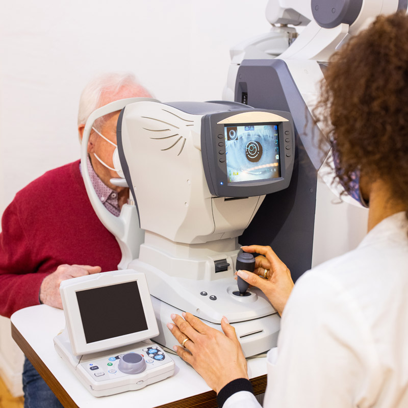
Image content: This image is available to view online.
View image online (https://my.clevelandclinic.org/-/scassets/images/org/health/articles/fundus-photography)
Fundus photography is a simple, noninvasive medical test where an eye care specialist takes a picture of the fundus, the back inner wall of your eye. These photos let your eye specialist take a close look at several parts of your eye that are very important to your vision.
Advertisement
Cleveland Clinic is a non-profit academic medical center. Advertising on our site helps support our mission. We do not endorse non-Cleveland Clinic products or services. Policy
Inside the back of your eye are several key structures. They are:
Fundus photography can be part of a routine eye exam. Your eye care specialist can also use it for diagnostic eye exams when you have eye symptoms or vision changes. Diagnostic eye exams look for more specific signs of eye diseases or conditions.
Some conditions or issues that might lead to fundus photography include:
Advertisement
Fundus photography involves using a fundus camera. Most eye care offices have at least one of these specialized devices.
There are two main types of fundus cameras:
Fundus photography doesn’t take any preparation on your part. Your eye specialist may give you eye drops to dilate your pupils before taking the photos. These drops take several minutes to start working.
There are two types of fundus cameras in widespread use. What you can expect depends on which camera type your eye specialist uses. In some cases, you may have pictures taken with both types.
For photos with these devices, you’ll sit on a chair or stool right in front of the frame at your end of the tube. Your eye specialist will use a sanitizing wipe to clean the forehead and chin rests before you put your face against them.
It’s important for your chin and forehead to be touching those rests. That makes it easier for your eye specialist to take the pictures more quickly. With your head in place, you’ll look into one end of the tube. Inside, you’ll see a light that’ll shine into your eye.
An eye specialist will sit on the opposite end of the tube. Looking into the camera’s eyepiece, the specialist will adjust the camera, aim it at specific points and take the pictures one at a time. They’ll do this for one or both eyes, depending on the reason for taking the pictures. Once they take the pictures, the test is done.
When you look into the wide-field camera opening, you’ll see and look directly at a circular target marker. The background behind the marker may change colors, and the eye specialist doing the test will tell you what the colors mean, how to move and anything else necessary to get the picture.
Once you’re in position, your specialist will activate the camera, and you’ll see a bright green light that sweeps across your vision like the light moving across a printed page inside a copier. It only lasts a second or so. They’ll usually take at least three pictures for each eye. Once they take the pictures, the test is done.
Advertisement
After taking fundus pictures, an eye specialist will probably take you to an exam room. In there, you’ll meet with an optometrist or an ophthalmologist. They’ll do any other parts of the exam that remain, or review your fundus pictures and talk to you about what they mean.
Fundus photography is an extremely safe test. It has no side effects. You may have side effects from medications for eye dilation (if you receive them), but these are usually mild and short-lived. If your eye specialist dilated your pupils, they’ll stay dilated until the medications wear off. Most of these medications wear off within a few hours (your eye specialist can tell you what timeframe is most likely for you, specifically).
If you have light sensitivity (photophobia), you might feel discomfort or find the lights that shine into your eyes during the photo-taking unpleasant. This usually only lasts a few moments and your eye care specialist can talk you through this. Most eye exam rooms also have dimmable lights, so your provider may use them if you’re still feeling discomfort from brighter light.
You may be able to drive without any difficulty after fundus photography. It depends on why you had the pictures taken, your eye condition before the test and other factors.
Advertisement
Most people who drive themselves to their eye specialist’s office can still drive after the test. If your specialist dilated your eyes, they may give you disposable shaded covers to wear that act like sunglasses. If you wear glasses, you can usually wear these disposable covers over your glasses until the dilating medications wear off.
Most eye care specialist offices have fundus cameras that can automatically upload photos to a computer in the exam room. Your eye specialist can pull the pictures up on a computer screen right then and there and check the pictures. They may also show you the pictures and explain the different things that you can see in the pictures.
If the fundus of your eye looks normal, your eye specialist will tell you that. If the pictures show any differences or changes that could be important, your eye specialist will explain them to you. If your eye specialist has previous pictures, they may pull up both sets and compare them. And depending on what they see in the most recent photos, they may do one or more of the following:
“A picture is worth a thousand words,” as the familiar saying goes. And a picture of the back of your eyes can be invaluable to your eye health. Your eye care specialist can take and use these pictures to check the health of your eye and diagnose any issues. And better still, fundus photography can help your specialist catch changes before they’re severe enough to cause symptoms. Fundus photography is part of routine eye exams, and your eye specialist can tell you more about how it can benefit you.
Advertisement


Sign up for our Health Essentials emails for expert guidance on nutrition, fitness, sleep, skin care and more.
Learn more about the Health Library and our editorial process.
Cleveland Clinic’s health articles are based on evidence-backed information and review by medical professionals to ensure accuracy, reliability and up-to-date clinical standards.
Cleveland Clinic’s health articles are based on evidence-backed information and review by medical professionals to ensure accuracy, reliability and up-to-date clinical standards.
Getting an annual eye exam at Cleveland Clinic can help you catch vision problems early and keep your eyes healthy for years to come.
