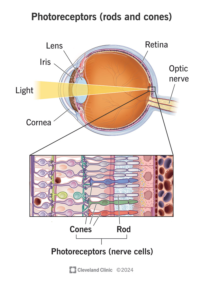
Lining your retinas are millions of special cells called photoreceptors. These cells are the key to turning light that enters your eyes into a form your brain can use for your sense of vision. Photoreceptors can be rod- or cone-shaped, and those shapes are part of how you can see in dim and bright conditions, and how you can see colors.
Advertisement
Cleveland Clinic is a non-profit academic medical center. Advertising on our site helps support our mission. We do not endorse non-Cleveland Clinic products or services. Policy

Image content: This image is available to view online.
View image online (https://my.clevelandclinic.org/-/scassets/images/org/health/articles/photoreceptors-rods-and-cones)
Photoreceptors (your rods and cones) are specialized light-detecting cells on the retinas at the back of your eyes. Their name comes from two ancient Greek words that combine to mean “light receivers.” They take light that enters your eyes and convert it into a form your brain can use for your sense of vision.
Advertisement
Cleveland Clinic is a non-profit academic medical center. Advertising on our site helps support our mission. We do not endorse non-Cleveland Clinic products or services. Policy
Your nervous system relies on cells called neurons, which use electrical and chemical signals to send and relay information. The photoreceptors in your retinas are a very specialized type of neuron. And because of how your retinas develop, they’re technically a part of your central nervous system (along with your brain and spinal cord).
Photoreceptors are a key part of how your eyes detect light and convert it into a form your brain can use. When you think about your eyes, it can be easy to think of them like cameras. But in reality, cameras — especially modern digital cameras — are technology based on how the human eye works.
Inside a modern digital camera, there’s a special sensor, and that sensor works very much like the human retina. It detects light and converts it into computer code. A tiny computer chip inside the camera then processes that code into a picture or video.
That’s basically the same process that happens when your retinas and brain work together so you can see the world around you. Your retinas turn what they detect into coded nerve signals and then send those signals through your optic nerve to your brain. Your brain decodes the signals and uses them to “build” what you see. And the cells in your retinas that start this process are the photoreceptors.
Advertisement
The human eye has two main types of photoreceptors, rods and cones, which get their names from their shapes.
These photoreceptors are tall and have a cylindrical (tubelike) shape. They’re extremely sensitive to even tiny amounts of light. About 95% of the photoreceptors in your eyes (about 100 - 125 million) are rods. They’re great at helping you see in dim places, but they aren’t as good at fine details, and they can’t see colors at all.
Rod photoreceptors are mainly responsible for low-light vision and night vision. When they help you see in dim light, that’s called scotopic (“sko-TOE-pick”) vision.
Cones are photoreceptors with a cone-like shape, meaning they’re circular at the bottom and have a pointed tip at the top. They need more light to activate than rods, but they can detect colors when they’re active. Most cones are in one place on your retina, the macula. They’re why the center of your visual field can see colors and other fine details.
Your eyes have three types of cones that work together as part of color vision. The cones don’t see the colors themselves. Instead, they see light wavelengths and tell your brain about them. Your brain turns that into your ability to see color.
When you look at a rainbow, you’re looking at a helpful way to understand the visible light spectrum. Red light has the longest wavelength, which is why it forms the outermost — and longest — part of the rainbow’s arc. Violet has the shortest wavelength, which is why it forms the inner — and shortest — part of the arc.
Most people have three types of cone photoreceptors. Having these three cone subtypes is called “trichromacy.”
The three subtypes are:
While the three cone types each specialize in a specific color, there’s a lot of overlap in sensitivity across the three types. Your brain can compare the differences in the signals from all three cone types to determine colors. That’s why the average healthy human eye can distinguish up to 1 million colors.
There’s a rare genetic mutation that can only happen in females that causes them to have four cone subtypes. That’s called “tetrachromacy” (tetra comes from the ancient Greek word for the number four). When tetrachromacy happens, it can be either weak or strong.
Weak tetrachromacy means the brain can’t fully process the input from the fourth cone. Strong tetrachromacy means the brain does process the fourth cone’s input. That fourth cone lets someone with tetrachromacy distinguish 100 million colors. But strong tetrachromacy is also very rare, which makes it hard for experts to tell how often it happens or do more research on it.
Advertisement
Several conditions can affect your rods and cones. Some of them are more general and affect other parts of the retina or surrounding eye tissue, too. But some conditions can affect your photoreceptors very specifically.
Conditions that can affect your photoreceptors include:
When you have a condition that affects your photoreceptors, vision loss is the main symptom. But that vision loss can take different forms, depending on the photoreceptors affected.
Many photoreceptor-related conditions affect both cones and rods at the same time. That’s especially true with conditions that involve damage or displacement of retinal tissue. Macular degeneration is a key, common example of that.
Advertisement
A standard eye exam is like an annual physical for your eye. It allows an eye care specialist to look at the back of your eye for any changes or other signs of tissue changes in your retina. Certain parts of the exam, especially eye dilation (mydriasis) and the slit lamp exam, can detect conditions that are otherwise invisible and don’t cause symptoms early on.
Other, more specific tests that might help include electroretinography (which measures electrical activity in your retinas) and visual evoked potentials (VEP). VEP is a brain test, but it can tell healthcare providers if signals from your retinas are reaching your brain.
And there are a few imaging tests that your eye care specialist may also recommend. These often involve seeing the different layers of the retina or mapping the blood vessels in and around your retinas. Doing that can help your eye care specialist see new blood vessel growth or other tissue changes that could lead to retinal damage or vision loss. Your eye care specialist can explain any additional testing options they recommend.
Your photoreceptors are part of your retinas, and taking care of your retina and eye health is key to maintaining your sense of vision. Some things you can do to take care of your retinas and eye health overall include:
Advertisement
When light lands on your rods and cones, it activates chemical and electrical processes in those receptors and the connected retinal cells. These processes are how the photoreceptor and retinal cells convert light into signals your brain can use.
Rods and cones are one of the most important parts of your sense of vision. Without them, your brain wouldn’t have the signals it needs to “build” what you see. And they specialize, too. Some let you see in the dark, while others let you see all the hues and shades the world around you has to offer.
Understanding how they work can help you do more than just appreciate their efforts. It can also help you maintain your eye health. If you have questions about your photoreceptors, retinas or other eye-related topics, an eye care specialist can help guide you. That way, you can maintain and get the most out of your sense of vision.

Sign up for our Health Essentials emails for expert guidance on nutrition, fitness, sleep, skin care and more.
Learn more about the Health Library and our editorial process.
Cleveland Clinic’s health articles are based on evidence-backed information and review by medical professionals to ensure accuracy, reliability and up-to-date clinical standards.
Cleveland Clinic’s health articles are based on evidence-backed information and review by medical professionals to ensure accuracy, reliability and up-to-date clinical standards.
Getting an annual eye exam at Cleveland Clinic can help you catch vision problems early and keep your eyes healthy for years to come.
