The retina is a key bridge between the light that enters your eyes and the images you see. Special cells in your retina react to light and pass signals to your brain that let you see the world around you. Talk to your eye care specialist if you notice gradual vision changes.
Advertisement
Cleveland Clinic is a non-profit academic medical center. Advertising on our site helps support our mission. We do not endorse non-Cleveland Clinic products or services. Policy
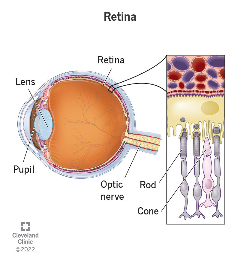
Image content: This image is available to view online.
View image online (https://my.clevelandclinic.org/-/scassets/Images/org/health/articles/22694-retina)
The retina is a layer of cells at the back of your eyeball that converts light into nerve signals. It then sends those signals along your optic nerve to your brain. Your brain processes those signals into your sense of vision.
Advertisement
Cleveland Clinic is a non-profit academic medical center. Advertising on our site helps support our mission. We do not endorse non-Cleveland Clinic products or services. Policy
Your retina senses light and sends signals to your brain. This happens because light-detecting cells called photoreceptors convert light into coded signals. Your brain receives those signals, decodes them and uses them to build the big-picture view you see with your sense of vision. That’s why retinal damage can change the way the world looks, leave gaps in your vision or cause total blindness.
Many conditions that can affect your retinas cause permanent damage and vision loss when not treated quickly. That’s why you should see your healthcare provider right away if your eyes or vision suddenly change.
Your retina is at the back of your eye, behind your iris and lens. When the focus is just right, the light rays converge just as they land on your retina.
Your retina has two main parts: your macula and your peripheral retina.
Many conditions that damage your eye can affect your retina. Retinal diseases include:
Advertisement
Talk to your healthcare provider if you notice any symptoms in your eyes, including:
Your eye specialist will check your retina as part of your overall eye exam. Your specialist will dilate your eyes for parts of this exam. Some retina-specific parts of the eye exam include:
Everyone needs to have their eyes examined regularly — at least every one to two years — whether or not you wear glasses or contacts. If you have certain chronic conditions that can affect your eyes, like diabetes, you need to see your eye care provider at least once a year. Your eye care specialist can recommend a schedule for regular eye exams if you need them more frequently.
There are several other precautions you can take to maintain and protect your eye health. They include:
See your healthcare provider as soon as you notice any changes in your vision. Whether it’s something as simple as needing new glasses or a more serious condition, don’t wait for symptoms to get worse before having your eyes checked.
Advertisement
Go to the emergency room if you have sudden vision loss or the sudden appearance of new vision changes or symptoms (sudden means they start or develop over minutes or a few hours).
These symptoms are possible with conditions that can quickly cause irreversible damage, leading to partial vision loss or blindness. You shouldn’t ignore them and should seek medical care right away.
Your retinas are a critical part of your vision, turning visible light into something your brain can process and work with. Without retinas, you couldn’t see the visible world around you. Taking care of your retinas is essential to protecting and maintaining your sense of vision. Talk to your provider about anything that seems “off” with your vision. The sooner you get a diagnosis and treatment, the more likely you are to avoid serious complications.
Advertisement


Sign up for our Health Essentials emails for expert guidance on nutrition, fitness, sleep, skin care and more.
Learn more about the Health Library and our editorial process.
Cleveland Clinic’s health articles are based on evidence-backed information and review by medical professionals to ensure accuracy, reliability and up-to-date clinical standards.
Cleveland Clinic’s health articles are based on evidence-backed information and review by medical professionals to ensure accuracy, reliability and up-to-date clinical standards.
Getting an annual eye exam at Cleveland Clinic can help you catch vision problems early and keep your eyes healthy for years to come.
