Retinal imaging is a diagnostic test that creates high-quality digital images of the inner, back surface of your eye. It allows the diagnosis of many eye conditions like diabetes-related retinopathy, glaucoma and macular degeneration. Your provider will tell you how often you need retinal imaging.
Advertisement
Cleveland Clinic is a non-profit academic medical center. Advertising on our site helps support our mission. We do not endorse non-Cleveland Clinic products or services. Policy
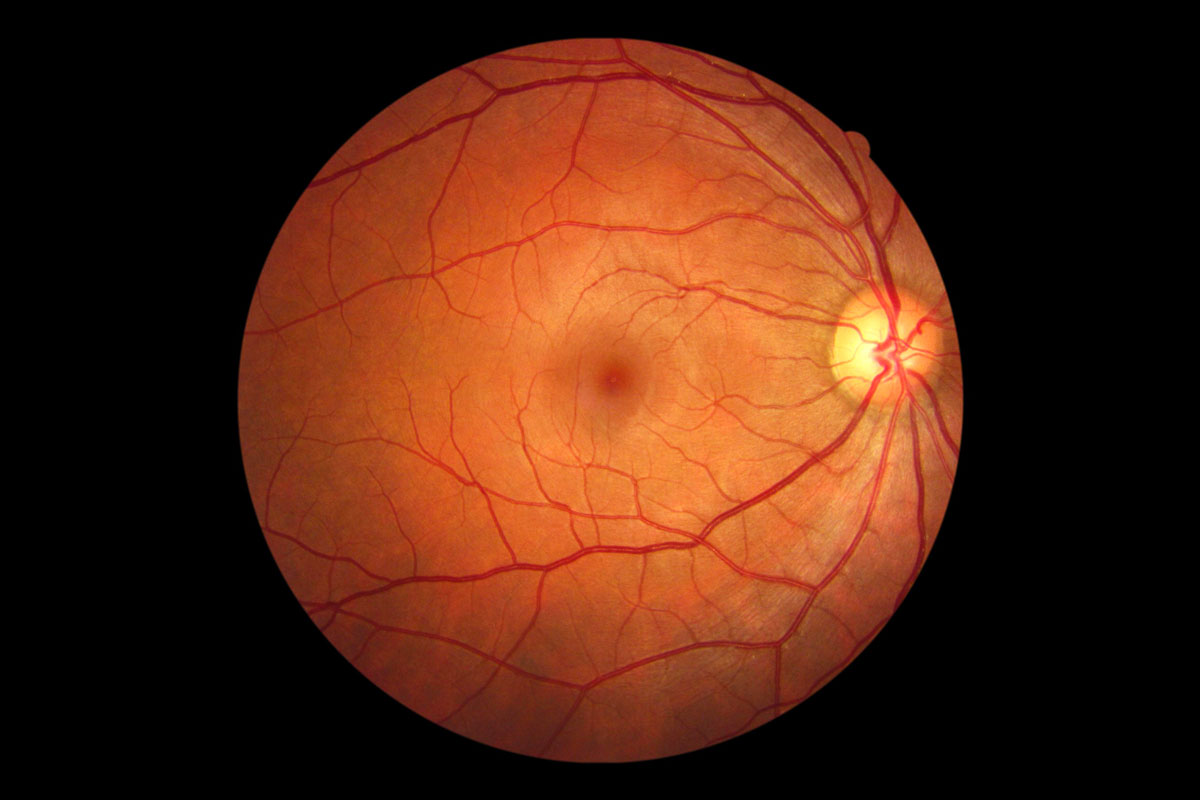
Image content: This image is available to view online.
View image online (https://my.clevelandclinic.org/-/scassets/images/org/health/articles/25046-retinal-imaging)
Retinal imaging refers to technologies your eye care specialist can use to create digital images of the inner, back surface of your eye. This part of your eye includes your retina (and an area within it called your macula) as well as your optic nerve and other important structures. Providers do retinal imaging as part of comprehensive eye exams and at follow-ups to monitor certain conditions.
Advertisement
Cleveland Clinic is a non-profit academic medical center. Advertising on our site helps support our mission. We do not endorse non-Cleveland Clinic products or services. Policy
Retinal images can show if you have certain eye conditions that, without treatment, could lead to vision loss. It helps to monitor how you’re responding to treatment. Repeated retinal imaging over many years allows your provider to notice subtle changes as they happen. Your provider will tell you what type of retinal imaging you need and how often.
Retinal imaging is a diagnostic test that can detect many different eye problems, including:
If imaging shows there’s an issue, your provider will recommend appropriate treatment. You may need serial imaging (photos of your retinas at regular intervals) to monitor your condition and see how well the treatment is working.
There are three main methods that eye care specialists use to take digital pictures of your eye’s fundus (the inner, back surface of your eye). These include:
Advertisement
In all these cases, you sit comfortably in a chair and move your face close to the camera device. Your provider shows you where to place your forehead and chin. Nothing touches your eye during retinal imaging.
Providers sometimes use one or more of the above methods at the same time. They may also use one of the methods along with fluorescein angiography. This technique is minimally invasive. Nothing touches your eyes, but your provider injects a dye into a vein in your arm. This dye travels through your blood vessels, including those in your eyes. It reveals blockages or other problems in those vessels.
Your provider will tell you about the procedure and why you’re having it. They may ask you to give consent.
Providers usually put drops into your eyes to dilate your pupils. That’s because your pupil is the window that the camera sees through to capture pictures of your fundus. A wider pupil typically leads to better images. Make sure you arrange for someone to drive you home. Your vision will be blurry, and you’ll be sensitive to light for a few hours after pupil dilation.
Retinal imaging, including fundus photography and OCT, is fast and painless. Here’s what you can expect:
Retinal imaging typically takes five to 10 minutes. If your provider uses fluorescein angiography, the process may take up to 30 minutes.
If your provider dilates your eyes, you’ll have blurry vision and sensitivity to light for a few hours. So, give your eyes a rest. Don’t drive, read or look at screens. Wear sunglasses to protect your eyes when you’re outside.
Retinal imaging, including fundus photography and OCT, is safe with no known risks. Your provider simply uses a camera to create digital images of your eyes.
Fluorescein angiography is low risk but may cause side effects like temporary skin discoloration (a slight yellow tint) or urine that appears dark yellow to orange. These effects go away within 24 hours.
Rarely, fluorescein may cause an allergic reaction (hives or itching). The risk of anaphylaxis is extremely small. People who are sensitive to fluorescein may experience:
This test produces digital retinal images that your provider will closely examine. Your provider may show you the images and explain what they mean. The images will become part of your medical record. Your provider can access them down the road as needed and compare them to newer versions to monitor changes in your eyes over time.
Advertisement
Call your provider if you have questions about what to expect at your appointment or what your test results mean. They’ll be happy to answer your questions and help you feel comfortable with the process.
Retinal imaging allows your provider to examine the inner, back surface of your eye. It’s vital for diagnosing and managing a range of conditions that can lead to vision loss without treatment. You don’t need to worry about anything touching your eye, and the whole process is quick and painless. Talk to your provider to learn more about the value of retinal imaging in your individual situation.
Advertisement


Sign up for our Health Essentials emails for expert guidance on nutrition, fitness, sleep, skin care and more.
Learn more about the Health Library and our editorial process.
Cleveland Clinic’s health articles are based on evidence-backed information and review by medical professionals to ensure accuracy, reliability and up-to-date clinical standards.
Cleveland Clinic’s health articles are based on evidence-backed information and review by medical professionals to ensure accuracy, reliability and up-to-date clinical standards.
Your eyes are one of your most important senses. If something goes wrong, it can change your world. Cleveland Clinic can help treat all types of retinal disease.
