A venogram (venography) is a test that uses X-rays to create moving pictures of blood flow in your veins, particularly those in your legs and pelvis. Providers use this test to diagnose deep vein thrombosis when ultrasound images can’t provide needed information. Venography also helps guide surgical procedures that treat vein disease.
Advertisement
Cleveland Clinic is a non-profit academic medical center. Advertising on our site helps support our mission. We do not endorse non-Cleveland Clinic products or services. Policy
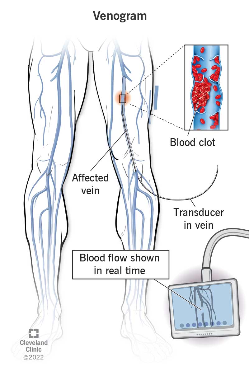
Image content: This image is available to view online.
View image online (https://my.clevelandclinic.org/-/scassets/images/org/health/articles/24929-venogram)
A venogram (venography) is an X-ray procedure that shows blood flow in your veins. Its main purpose is to help healthcare providers diagnose and manage conditions that affect your veins, including blood clots.
Advertisement
Cleveland Clinic is a non-profit academic medical center. Advertising on our site helps support our mission. We do not endorse non-Cleveland Clinic products or services. Policy
In many cases, providers use vascular ultrasound to diagnose blood clots in your veins. However, venography is an alternative when ultrasound isn’t possible or doesn’t provide enough information. Providers also use venography to plan or guide venous disease treatments.
A venogram can evaluate blood flow in many different veins throughout your body. Providers typically use this test to look at veins in your:
Unlike ultrasound, a venogram is invasive. That means a provider has to make a small needle puncture in your skin to access your veins. They inject a contrast dye into your veins so they can see your blood flow on the X-ray. Since the test uses X-ray technology, it involves a small dose of radiation.
Most people experience only mild discomfort during the test, and they have no serious side effects. However, this test may not be safe for some people, including those who are pregnant. It’s important to talk to your provider before having a venogram. Your provider will decide if the test is safe and appropriate for you.
The medical term “venography” refers to the testing process, while “venogram” refers to the resulting images. But people usually use “venogram” to mean both the test and the images.
Advertisement
Providers may order a venogram to:
Venography uses X-rays (a form of radiation) to capture images of the inside of your body. Because veins normally don’t show up on an X-ray, your provider injects a contrast dye into your veins. This dye flows through your veins and makes them visible, allowing your provider to see blood clots or other blood flow problems.
While X-rays capture single moments in time, a single moment isn’t enough to show blood flow, which is a dynamic process. This means that providers need to see blood moving through your veins while it’s actually happening. So, to generate venograms, they use a technique called fluoroscopy. This method uses several pulses of an X-ray beam to take continuous, real-time images.
Think of the many individual pictures that come together to make an animation. That’s similar to how a venogram works. The moving pictures allow your provider to see your blood flow in action.
Your provider will tell you how to prepare. They may ask you to fast (have no foods or drinks except water) for a few hours before the test. They’ll also let you know if you need to stop taking any medications.
Be sure to tell your provider:
Before the test begins, you’ll need to remove jewelry and any metal objects that could disrupt the X-ray process. You should wear loose, comfortable clothing, but your provider may give you a gown to wear.
Yes, you’re typically awake for a venogram. Your provider may give you a sedative to help you relax.
A radiologic technologist performs this procedure. You can expect them to do the following steps:
Advertisement
There may be additional steps depending on the reason for your venography. For example, if venography helps guide a procedure (like thrombolytic therapy), your care team will perform other steps to give you the treatment you need. Your provider will explain what you should expect in your individual situation.
Venography typically takes 30 to 90 minutes.
Your provider will briefly monitor you for allergic reactions or other complications. They’ll tell you when you can go home. They may ask you to drink plenty of water for the next 24 hours to help flush the contrast dye out of your body.
Risks of venography include:
Usually, the benefits of venography outweigh the possible risks. In some cases, venography may be too risky for you because of an underlying medical condition (like severe kidney disease). Discuss all possible risks with your provider before the procedure.
You may feel sick to your stomach or a brief flushing sensation when your provider injects the contrast dye. That should quickly pass.
Rarely, people experience a delayed reaction to the contrast dye that begins hours or days later. This reaction is usually mild and can include:
Advertisement
Moderate side effects may include:
Severe side effects include:
Call your healthcare provider right away if you have any delayed reactions. If your side effects are severe, seek emergency care. They could be signs of a serious allergic reaction that needs immediate treatment.
Venography produces images that a radiologist will analyze. They’ll send a report to the provider who referred you for the test. Your provider will then talk with you about the results and explain what they mean. For example, the images may show you have a blood clot in a deep vein in your leg. Your provider will then recommend treatment based on the findings.
Sometimes, providers use venography for real-time imaging guidance during surgical procedures. In that case, your provider may not discuss results with you. Instead, they use the images in the moment to guide your treatment. They’ll let you know if any issues arise.
Call your provider if you:
Advertisement
Both procedures show blood flow through your blood vessels using X-ray technology and contrast dye. The difference is that a venogram specifically evaluates your veins. An angiogram evaluates arteries or veins. Healthcare providers consider angiograms the gold standard for finding blockages in your arteries. They sometimes give you treatment (like angioplasty) at the same time.
Providers use venograms less often for diagnostic purposes, instead preferring other methods (like ultrasound) to find blockages in your veins.
Medical testing can be stressful. You may worry about discomfort during the test or what the results will show. Be assured that your healthcare provider is there to answer your questions. Don’t hesitate to share your concerns and get the information you need to feel more comfortable with the process.


Sign up for our Health Essentials emails for expert guidance on nutrition, fitness, sleep, skin care and more.
Learn more about the Health Library and our editorial process.
Cleveland Clinic’s health articles are based on evidence-backed information and review by medical professionals to ensure accuracy, reliability and up-to-date clinical standards.
Cleveland Clinic’s health articles are based on evidence-backed information and review by medical professionals to ensure accuracy, reliability and up-to-date clinical standards.
Vascular disease may affect your life in big and small ways. Cleveland Clinic’s specialists treat the many types of vascular disease so you can focus on living.
