X-rays are a way for healthcare providers to get pictures of the inside of your body. X-rays use radiation to create black-and-white images that a radiologist reads. Then, they send a report to your provider. X-rays are mostly known for looking at bones and joints. But providers can use them to diagnose other conditions, too.
Advertisement
Cleveland Clinic is a non-profit academic medical center. Advertising on our site helps support our mission. We do not endorse non-Cleveland Clinic products or services. Policy
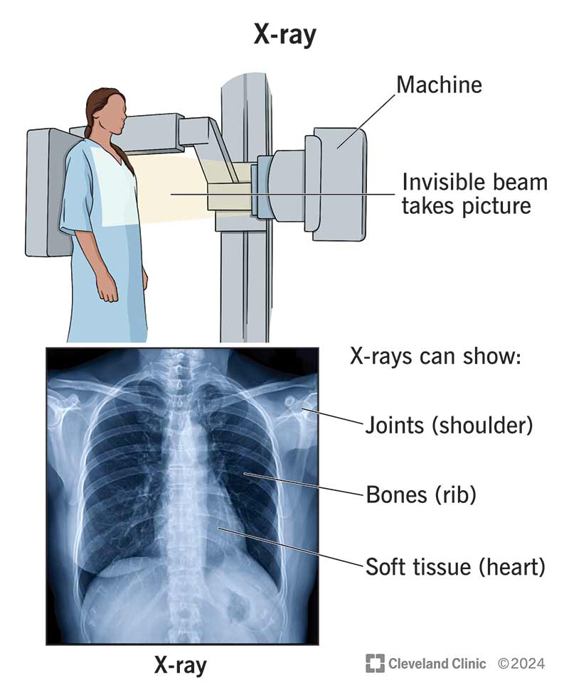
Image content: This image is available to view online.
View image online (https://my.clevelandclinic.org/-/scassets/images/org/health/articles/21818-x-ray)
An X-ray is a type of medical imaging that uses radiation to take pictures of the inside of your body. We often think of an X-ray as something that checks for broken bones. But X-ray images can help providers diagnose other injuries and diseases, too.
Advertisement
Cleveland Clinic is a non-profit academic medical center. Advertising on our site helps support our mission. We do not endorse non-Cleveland Clinic products or services. Policy
Many people think of X-rays as black-and-white, two-dimensional images. But modern X-ray technology is often combined with other technologies to make more advanced types of images.
Some specific imaging tests that use X-rays are:
X-rays can help healthcare providers diagnose various conditions in your body. Some of the most common areas on your body to get an X-ray are:
Advertisement
X-rays work by sending beams of radiation through your body to create images on an X-ray detector nearby. Radiation beams are invisible, and you can’t feel them.
As the beams go through your body, bones, soft tissues and other structures absorb radiation in different ways:
A radiologist interprets the image and writes a report for the physician who ordered the X-ray. They make note of anything that’s abnormal or concerning. Then, your healthcare provider shares the results with you.
Preparation for an X-ray depends on the type of X-ray you’re getting. Your provider may ask you to:
Tell your healthcare provider about your health history, allergies and any medications you’re taking. Let them know if you’re pregnant or think you could be.
The exact steps of an X-ray depend on the kind you’re getting. In general, your provider will follow these steps during an X-ray:
Sometimes, children can’t stay still long enough to produce clear images. Your child’s provider may recommend using a restraint during an X-ray. The restraint helps your child stay still and reduces the need for retakes. The restraints don’t hurt and won’t harm your child.
Most of the time, there aren’t any restrictions on what you can do after an X-ray. You can go back to your typical activities.
X-rays are safe and low risk.
X-rays use a safe and small amount of radiation — not much more than the naturally occurring radiation you get in your daily life. For instance, a dental X-ray exposes you to about the same amount of background radiation you’d get in one day.
X-ray radiation is usually safe for adults. But it can be harmful to a fetus. If you’re pregnant, your provider may choose another imaging test, like ultrasound.
Advertisement
A radiologist will review your X-ray and take notes of what they see and what they recommend. Your healthcare provider will receive both the images and the radiologist’s notes. They’ll review the images, too, and let you know if they see anything concerning.
It depends on when the radiologist can look at them. You typically get X-ray results back much more quickly if you’re in an emergency. Results from a bone X-ray are often ready right away, but it could take several minutes or hours for a radiologist to read and record the results. Results from more complex X-rays may take longer.
Your provider might share your results with you after the X-ray, or you might see them show up in your electronic health records. Talk to your provider about when you can expect your results.
It depends on what the findings are and if it’s harmful. Your provider will let you know if you need more imaging or testing to take a better look at something. They could also refer you to a provider who specializes in a certain organ (like a pulmonologist for the lungs). Sometimes, they recommend a follow-up X-ray to see if a finding changes over time.
Check with your provider if you have any questions about the X-ray results.
Advertisement
X-rays can show cancer, but it’s not how providers look for or diagnose most cancers. This is because tumors in your organs can be small or hidden behind other structures in your body, or they can blend in with normal tissues.
X-ray is one of the oldest, most reliable medical technologies. And despite it being almost 130 years since its discovery, it’s still relevant thanks to research into new, better ways to use it. Modern X-rays are far more detailed and use less radiation than they did in the past, thanks to advances in imaging resolution.
X-rays allow providers to quickly check what might be going on inside your body. This means you can know very soon whether there’s something that needs treatment or additional testing. Let your provider know if you have any questions about getting an X-ray or what your results mean.
Advertisement


Sign up for our Health Essentials emails for expert guidance on nutrition, fitness, sleep, skin care and more.
Learn more about the Health Library and our editorial process.
Cleveland Clinic’s health articles are based on evidence-backed information and review by medical professionals to ensure accuracy, reliability and up-to-date clinical standards.
Cleveland Clinic’s health articles are based on evidence-backed information and review by medical professionals to ensure accuracy, reliability and up-to-date clinical standards.
When you need a clear picture of what’s happening inside your body, the Cleveland Clinic imaging team is here for you.
