Locations:

Echocardiogram: Transesophageal (TEE)

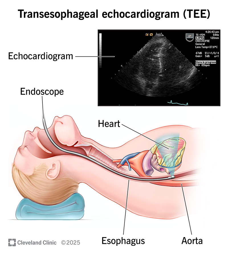
A transesophageal echocardiogram (TEE) is a type of echo test that uses sound waves to create pictures of your heart. Unlike other types of echo tests, a TEE creates pictures from inside your body. Your provider guides a thin, flexible tube down your esophagus. A TEE can diagnose many problems including blood clots and infections in your heart.

Overview

What is a transesophageal echocardiogram?

A transesophageal echocardiogram (TEE) is a test that uses sound waves to create pictures of your heart. A TEE is different than other forms of echocardiogram or ultrasounds because it takes pictures from within your body, rather than outside of it.

Advertisement

Cleveland Clinic is a non-profit academic medical center. Advertising on our site helps support our mission. We do not endorse non-Cleveland Clinic products or services. Policy

Your provider uses a long, thin tube called an endoscope to carefully guide a small transducer down your throat and esophagus (food tube). The transducer is a device that makes sound waves. These sound waves bounce off the different areas of your heart, making echoes. The transducer then sends these echoes to a computer that makes them into pictures. These pictures show the structure and function of your heart in great detail.

Your provider may combine TEE with Doppler ultrasound and color Doppler methods. These specialized techniques show the speed and direction of blood flow through your heart.

What does a transesophageal echocardiogram show?

A transesophageal echocardiogram shows a detailed view of your heart’s structure and function. It can help diagnose and manage many different conditions, including:

  • Aortic aneurysm, which is a bulge in your largest blood vessel.
  • Blood clots in your heart. People with atrial fibrillation face a higher risk of clots in their atria (upper heart chambers).
  • Cardiac tumors, which may be cancerous or noncancerous.
  • Congenital heart disease, which includes a range of heart problems that you’re born with.
  • Heart valve disease, including mitral valve disease. Your valves may be narrowed (stenosis) or leaky (regurgitation). TEE can also spot problems with prosthetic valves, such as growths or infections.
  • Infective endocarditis and other infections in your heart tissue or valves.
  • Pericardial disease, which affects the sac that covers your heart.

Advertisement

Why is a transesophageal echocardiogram performed?

A transesophageal echocardiogram may be performed for many reasons. These include:

  • You had an echocardiogram on the outside of your chest (a transthoracic echocardiogram or TTE), but your provider needs a better or more detailed visualization of your heart. In this case, a TEE is a follow-up to the TTE that helps diagnose or manage a heart problem.
  • You have a life-threatening problem that your provider needs to see up-close, right away. TEE can be useful in emergency and critical care.
  • Your provider needs to check for blood clots before you have a medical procedure such as cardioversion.
  • You’re having a surgery or procedure. TEE helps providers confirm your surgery was successful. TEE also provides real-time imaging during some catheter-based procedures.

Diagnosing or managing a heart problem

Your provider may need more information than what a TTE, performed outside your body, can provide. So, they may run a TEE to examine your heart without your ribs or lungs getting in the way.

Imagine standing on a sidewalk, looking through a storefront window. Inside the store, near the back, you see a glass display case. You can tell the display case contains plates with flower patterns. But you can’t make out the details since people are standing around the case. So, you go inside and walk to the back of the store to get a better look.

A transthoracic echo gives your provider a storefront window view, which is still very helpful. But a transesophageal echo lets your provider see directly inside the display case. It’s the difference between seeing the plate has flowers and learning the flowers are roses.

Since your esophagus runs behind your heart, it’s the perfect spot for the transducer to send and receive sound waves. A TEE is especially valuable for examining structures in the back of your heart. These include your:

  • Left atrium: The top left chamber of your heart.
  • Left atrial appendage (LAA): The small, ear-shaped sac in your left atrium where blood clots sometimes form.
  • Interatrial septum: The wall that separates your left atrium and right atrium.
  • Thoracic aorta: The part of your aorta that passes through your chest.

Sometimes, providers skip the TTE and use TEE first. This may be the case if you have:

  • Chest anatomy that makes a transthoracic echo difficult.
  • Lung disorders.

Supporting emergency and critical care

TEE can help investigate life-threatening medical conditions. In some critical situations, providers use TEE without using TTE first. This may happen if you have one of the following conditions without a clear cause:

  • Hemodynamic instability (problems with blood pressure and blood flow).
  • Hypotension (low blood pressure).
  • Hypoxia (low oxygen).

Advertisement

TEE may also be used initially, without a prior TTE, to investigate:

  • Aortic dissection or other sudden problems with your aorta that may require urgent investigation.
  • Heart attack.
  • Prosthetic valve problems (like a blood clot or infection).
  • Trauma to your chest.

Checking for blood clots before a procedure

Providers use TEE to check for blood clots before certain procedures that treat arrhythmias. These include:

These procedures may cause a blood clot to break free and travel through your bloodstream (thromboembolism). Your provider will let you know if you need TEE to check for clots before your procedure.

Helping with surgeries and procedures

TEE is a valuable tool during certain surgeries and procedures. Your provider may use TEE before your surgery to check your anatomy. They may also use TEE afterward to confirm the surgery’s success. TEE may be used for:

  • Surgery for congenital heart disease.
  • Valve repair.
  • Valve replacement.

Providers also use TEE to see inside your body and guide procedures in real time. TEE may be used during percutaneous procedures including:

Is TEE an invasive procedure?

A TEE is an invasive procedure because your provider needs to put a device inside your body. You may experience mild discomfort. But the risk of serious problems is low.

Advertisement

Does TEE require anesthesia?

Usually, a TEE requires only local anesthesia and moderate sedation. That means your provider will spray the back of your throat with pain-relieving medication. They’ll also give you medicine through an IV to help you relax.

Most people don’t need general anesthesia (deep sleep). Talk with your provider about what’s best for you during your TEE.

Are you awake during a transesophageal echocardiogram?

Moderate sedation should make you drowsy and allow you to tolerate the procedure without significant discomfort. The staff will be constantly monitoring for signs of discomfort and can decide if you need more sedation. . If you need general anesthesia, you’ll be fully asleep and you won’t remember the procedure. With general anesthesia, you will need to be placed on a breathing machine. Because you are deeply sedated, the recovery will be longer compared to moderate sedation.

How long does a transesophageal echocardiogram take?

A TEE examination may take up to 90 minutes. The picture-taking part usually only takes 15 minutes.

Test Details

How do I prepare for a transesophageal echo?

Talk with your provider about your medical history. Some medical conditions make a TEE too risky to perform. Tell your provider if you have:

Advertisement

Also tell your provider if you take medication for:

Your provider will give you detailed instructions on how to prepare for your TEE. Be sure to follow them closely and ask questions if anything is unclear. These instructions will tell you:

  • When to stop eating and drinking. This will usually be at least six hours before the test.
  • When to take your usual medications.
  • When to avoid drinks containing alcohol. You may need to avoid them for several days leading up to your test.
  • Anything else you should do or avoid before your TEE.

What can I expect during a transesophageal echo?

A transesophageal echo includes the following steps:

  1. You’ll put on a hospital gown.
  2. Your provider will place electrodes (small stickers) on your chest. The electrodes allow your provider to watch your heart’s electrical activity during the TEE.
  3. Your provider will place a blood pressure cuff on your arm, and a pulse oximeter on your finger (to check your oxygen level).
  4. You’ll gargle with a solution to numb your throat. Your provider will also spray your throat with medication. This is a local anesthetic that prevents you from feeling any pain during the TEE.
  5. You’ll receive a sedative through an IV. This will help you relax.
  6. You may have a tube in your nose that provides oxygen called a nasal cannula.
  7. You’ll lie on your left side on the exam table.
  8. The provider will gently insert an endoscope into your mouth. This is a long, thin, flexible tube. It has a transducer on its tip. The tube travels into your throat and esophagus. It’s lubricated to help it slide down more easily. Your provider may tell you to swallow at certain points to help the transducer get into the proper position.
  9. Your provider will take pictures. You shouldn’t feel any pain. But you may feel a bit of pressure in your chest as your provider moves the transducer into different positions. This part will take about 10 to 15 minutes.
  10. Your provider will remove the tube from your throat. They’ll continue to watch your blood pressure, pulse and oxygen level.
  11. Your provider will tell you when it’s OK to leave.

What can I expect after a transesophageal echo?

Someone will need to drive you home from your TEE. You won’t be able to drive for 24 hours after your TEE due to the sedation. You can expect to:

  • Have a numb throat for an hour or so. This will wear off. Don’t eat or drink anything until the numbness goes away. You might choke if you eat or drink too soon.
  • Have a sore throat for up to two days. This is normal and will go away. If it doesn’t go away or gets worse, call your provider.

What are the risks of a transesophageal echo?

Some people have a sore throat after a TEE. It may last a few hours or up to three to five days.

Other complications are less common but may include:

  • Allergic reactions to medications.
  • Aspiration pneumonia.
  • Blood pressure or heart rhythm problems.
  • Minor bleeding in your esophagus.

Talk with your provider about the risks of a TEE before your procedure.

Results and Follow-Up

How do I get the results of my TEE?

Your provider will discuss your test results with you. They may also be available as part of your electronic medical record (an online resource that lets you see your medical information). Talk with your provider about when your results will be available and how you can view them.

A note from Cleveland Clinic

Medical tests make some people feel nervous or uneasy. It’s normal to feel that way. But tests like a transesophageal echocardiogram play a huge role in helping you stay healthy or treating problems that come up. A TEE is a common procedure that has a low risk of complications. If you need a TEE, talk with your provider about how to prepare and what to expect.

Medically Reviewed

Last reviewed on 05/06/2022.

Learn more about the Health Library and our editorial process.

Ad
Appointments 800.659.7822