Cleveland Clinic’s Endoluminal Surgery Center and Lesions in the Lower Tract

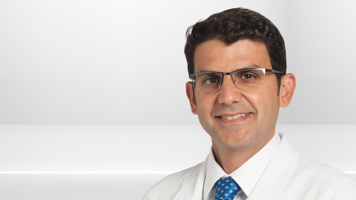
Emre Gorgun, MD, colorectal surgeon and co-director of Cleveland Clinics Endoluminal Surgery Center, joins the Cancer Advances podcast to discuss the center and management of lesions in the lower GI tract. Listen as Dr. Gorgun explains our approach to resecting lower GI polyps and how this allows patients to avoid surgery, keep their native organs and have an improved quality of life.
Subscribe: Apple Podcasts | Buzzsprout | Spotify
Cleveland Clinic’s Endoluminal Surgery Center and Lesions in the Lower Tract
Podcast Transcript
Dale Shepard, MD, PhD: Cancer Advances, a Cleveland Clinic podcast for medical professionals, exploring the latest, innovative research and clinical advances in the field of oncology. Thank you for joining us for another episode of Cancer Advances. I'm your host, Dr. Dale Shepard, a medical oncologist here at Cleveland Clinic overseeing our Taussig Phase I and Sarcoma Programs. Today, I'm happy to be joined by Dr. Emre Gorgun, a colorectal surgeon and co-director of the Cleveland Clinic Endoluminal Surgery Center. In another episode of this podcast, I talked to Dr. Bhatt about management of lesions in the upper GI tract. Dr. Gorgun is here today to talk to us about lower GI surgeries. Welcome.
Emre Gorgun, MD: Thank you very much, Dale, and thank you for having me here for the second time. It's really an honor to be chatting with you.
Dale Shepard, MD, PhD: Absolutely. I'll comment that you have another episode where we talked about organ-sparing approaches to rectal cancer, but today we're going to talk about endoluminal surgeries and the Endoluminal Surgery Center. So maybe we can just take a really brief step back, and let the listeners know what exactly is endoluminal surgery.
Emre Gorgun, MD: Thank you, Dale. Really a great question because sometimes it can be a little confusing. What does that really mean? What is endoluminal surgery?
I guess we can start by not to lecture here or anything like that, but what we mean by that is lumen is an organ, a hollow organ, or like an intestine and doing a surgery, staying inside of that intestine is, in basic terms, what we mean by that. I mean, this is more of a space or specialty within our group or gastrointestinal surgery or gastrointestinal diseases. It's still evolving and developing, but it is a very exciting space. And it truly help our patients greatly in terms of, again, organ sparing, to not to necessarily take out a major part of patient's organs, or intestines, or colons, or rectums, or even stomach as my co-director, Dr. Bhatt will be talking in a different podcast.
So what we mean by that is there's a large number of patients discovered on a yearly basis with abnormal tissues in the colon and rectum, and some of them are really early lesions, thanks to our widespread screening protocols and screening programs within the United States. As you know also the Preventive Task Force in the United States recently increased the age cut-off for screening to 45. With that, we expect to see even more of these new pre-malignant or early malignant lesions in the colon and rectum. Of course, I'm mostly speaking specifically for the lower GI, which means our colon and rectum, the last portion of our intestinal tract before it exits from the anus.
So with these screening colonoscopies, when the lesions are discovered, it's unfortunately a common referral pattern within the United States that these patients are typically referred to surgeons like myself, and like my colleagues, colorectal surgeons, for removal of these lesions because these lesions, the majority of the time been unamenable to be removed endoscopically. However, advanced endoscopist and surgical endoscopist or gastroenterologist we believe who are really passionate about this approach is that without doing surgery, we can take these lesions out endoscopically. But as we have been doing these procedures more and more, we developed our techniques, we developed our approaches, we improved our skills as proceduralists, as well as our tools and instruments.
Devices are also refined and improved to the point that compared to what we used to do, maybe 10, 15, 20 years ago, now we can really do more technically challenging procedures and tissue removals, these abnormal tissue removals inside of the intestine to the point that recently we started to call this approach endoluminal surgery, because it is not necessarily just the putting a snare or lasso around the polyp, like a mushroom type of lesion and just cutting it off or chopping the lesion off. It's more than that. We really precisely take these lesions millimeter by millimeter and remove it from the underlying muscle layers of the intestinal wall, that safely we can remove these lesions and still get good pathology analysis, as well as preserve someone's organ.
That's why this is an area and space that I'm privileged to share with you that we started to call this with few other physicians in the United States, but definitely take pride in that we probably are one of the first endoluminal surgery centers, if not the first, that has this type of a center and offers this type of services to our patients.
Dale Shepard, MD, PhD: When we think about endoluminal surgery compared to the traditional surgery, or, as you say, oftentimes in a colonoscopy, they will just try to pluck out a polyp, what would be an ideal lesion that should not be removed during the colonoscopy, but be referred for an endoluminal procedure? Is there a location, is there a size? What are the criteria that you would like to see in terms of who comes to you for this procedure?
Emre Gorgun, MD: Speaking for the lower GI, again, I'm going to concentrate on the colon and rectal lesions, and that's how we divided our work among ourself within our center, upper and lower, I'm happy to share with you that any location within the colon and rectum is applicable for this type of approach.
In lesions that are complex there are, obviously, the majority of the smaller polyps, and these are usually 85% of the lesions that are discovered during regular screening colonoscopy, are able to be removed by endoscopist that is not necessarily trained or skilled in advanced endoscopy. So majority of these smaller polyps can be snared, removed with forceps or with snares different sizes.
However, when it comes down to these 15% of lesions, it can be anywhere in the colon, can be in the rectum, but typically to the eyes of the endoscopist, strictly endoscopist, quote-unquote, these can be deemed not easily removable by their assessment, and these lesions can be then sent to surgeons. So these are the ones that we talking about, little bit more complex. They're definitely not easy to be removed, looks larger, a little bit larger and the society guidelines usually indicate definitely larger than 2 centimeters.
But having said that, there are some lesions that are smaller, but they are a little bit more complex. Either they are scarred or puckered in the middle. They might be some feature. So I don't want to limit it to necessarily just the size, but complex is, I think, a better term that we can describe.
But one way or another, the primary screening endoscopist does not feel like these lesions are easy. So these certainly are good candidates for our center to be patients and to be possibly considered for endoluminal surgical removal.
Having said that, of course, if there's clear-cut pathology with biopsy that reveals cancer, more than like an early cancer, but certainly a large mass, these are, of course still... I need to mention that these need to be removed surgically, of course.
Dale Shepard, MD, PhD: So, is there an educational component here perhaps that people doing the primary colonoscopies might be aware that this is a potential, so they either don't attempt to remove something maybe they shouldn't or make the right referral patterns? And if so, how does that happen?
Emre Gorgun, MD: Hundred percent. This is such a good point, and thank you for, Dale, bringing this up. This is also in the guidelines of different societies as well.
Certainly a great amount of education goes into referring physician. Publications or activities such as this are very important from that perspective, and we try to send out newsletters, information, bulletins as well as publications, of course, scientific publications are towards that, but society guidelines are out there as well. But still that's a continuous education and communication between us and the referring physicians. That's why always I prefer to communicate back to our referring physicians about this type of details.
So what are education? Certainly recognizing the lesions. Possibly if something looks non-malignant, not necessarily biopsying it because when you biopsy these lesions that can create more scarring in the lesion and can actually make our job for endoluminal surgery a little bit harder. Not necessarily putting a tattoo. When endoscopists find these lesions, they tend to put a blue dye or a dye to mark that lesion so when someone comes back can easily identify these. But putting these dyes right under the lesion is not a good practice as well because that can create also scarring of the lesion down to the deeper tissues and can make our procedure, endoluminal procedures, harder. So these are definitely important educational points that we like to inform our referring physicians.
Dale Shepard, MD, PhD: You mentioned before about size, or you mentioned complexity of things that may be should come to you when you're actually doing the procedures. Is there a size or complexity where it's not appropriate to do this? What are those kind of boundaries, and when would you decide not to do an endoluminal procedure and go to surgery?
Emre Gorgun, MD: Size-wise, certainly I can't tell you that there's a upper limit and there's certainly not a lower limit. Obviously, the smaller lesions takes less time to completely remove them. But, again, some small lesions can be harder as well because of different reasons like scarring, puckering, depression down to the attachment to the muscle layer for different reasons, either from previous procedures or even maybe an early cancer. These can be difficult.
Having said that, when we look at this different studies, the fact that a lesion is large does not necessarily always indicate that it has a higher risk for malignancy or cancer, so that's a good news. Sometimes really benign lesions, non-cancerous lesions, can grow really big sizes and what we call them more like carpeted lesions covering a large surface area, even sometimes a circumferential lesion that is encompassing the entire circumference of the lumen. So these can be even benign. I've done multiple of these lesions, even sometimes it's very satisfying. We can even remove these circumferential lesions in an en bloc fashion, almost like a cylinder and remove the entire intestinal lining and the pathology can be still non-cancer. So size-wise, there's nothing that would necessarily stop us.
But the pit patterns or what we call it, or the anatomy like surface anatomy, how it looks from outside gives us a lot of information in terms of determining whether a lesion is malignant or benign. So from that perspective, certainly there are factors that we weigh in or make decisions upon.
Dale Shepard, MD, PhD: When we think about lower GI cancers, we think about colon cancers as a hereditary component. One of the strengths here at Cleveland Clinic is certainly the Weiss Center. What sort of collaboration do you have between the Weiss Center and hereditary cancers and these endoluminal procedures?
Emre Gorgun, MD: We have a very strong collaboration and link to our Weiss Center and Hereditary Colorectal Cancer Group. We work with them extremely closely. By the nature of what we do and what we treat here under our endoluminal surgical center umbrella, we do see a lot of patients that are candidates for further workup and get them connected with our Hereditary Colorectal Cancer Group. We are privileged to have a large group of providers within that group that provides us, helps us with genetic counseling and information.
Very frequently also as Endoluminal Surgery Center, we receive patients that have also not only one polyp, but several polyps throughout their colon at different age groups or with a strong family histories, et cetera. We have a care path that we channel them into this Weiss Center as well so we can get their genotyping and panel there and genetic profiling and include them into our registry within the Weiss Center. So there's certainly a great collaboration and we are, like I said, privileged and in a unique position to be able to provide or offer our patients these type of services as well.
Dale Shepard, MD, PhD: When we think about outcomes, tell us a little bit about outcomes. Traditionally, we sort of think of this as colonoscopy, remove some polyps, or you find a tumor, go to surgery. This is a totally different way to think about how to manage polyps and sort of precancerous positions and repeat colonoscopies. What kind of outcome data do we have in terms of our ability to prevent cancers?
Emre Gorgun, MD: Yes, that's something that we really paid attention on it. And with our care coordinators and program managers within our group, we were privileged to work with outstanding providers, and we developed our care paths.
With that, certainly when a patient is referred to us to our center, we follow these care paths very closely. Certainly due to the reasons we discuss before, we need to decide by looking at the anatomy or surface, the specific features of the endoscopic view, first step is, is this a good match, good patient for our center or not? Can we really approach these patients first with endoluminal surgery? For that, certainly colored images is the first thing that we request and to understand better what the specifics of these lesions are because really pictures will mean much more than a paragraph, even, at some cases. And as humbly speaking, having trained eyes and visual feedbacks in our minds, we can make a lot out of these colored images.
Based on that, we then decide whether we can treat this under general anesthesia settings or under conscious sedation settings so we have the ability to offer our patient services, both inpatient and outpatient, and then these procedures are performed. Following to that, certainly there's a close follow-up protocols that we also incorporate it into our care path, and certainly all advanced endoscopy or all endoluminal surgical patients are again surveyed with colonoscopy in six months' period to make sure what the outcomes or what their final results are.
So that brings us back to your question, what are the outcomes? Certainly what we are monitoring there is, do we have any high recurrence rates? Yes, we think that we are removing these lesions, but are these lesions are completely eliminated from the colon surface, from the rectal surface, or do they tend to come back? So in our registry, in our follow-up outcome books, outcome registries, I'm very happy to share with you is that our recurrence rates for these polyps are less than 2% and that's way below the national averages.
When they do come back, they are in the form of adenomas and non-malignancies, and 99%, we can still remove these endoscopically with shorter endoluminal surgical approaches. So, ultimately, there are very, very rare incidences that we need to do surgically take these lesions out, either taking part of their colon or rectum, but I'm happy to share with you that majority of these lesions are managed purely endoluminal at the end.
Cancer risks are also very low. Of course, if you find a cancer then in the final pathology that requires more advanced, more oncological resection, also that puts us in a unique position as well, that as surgeons, as colorectal surgeons, then we can streamline these patients very quickly and get their colon resections or rectal resections scheduled very efficiently with the help of our outstanding teams and provide them the best healthcare that our patients deserve.
Dale Shepard, MD, PhD: Well, that's really fascinating how we've been able to develop this, appreciate all of your efforts to do that. This is a great option for patients, and I appreciate your insight.
To learn more about Cleveland Clinic's Endoluminal Surgery Center, or to refer a patient, please call (216) 444-1244. That's (216) 444-1244. You can also visit the website at clevelandclinic.org/els. That's clevelandclinic.org/els. Thank you very much again.
Emre Gorgun, MD: My pleasure, Dale. Thank you very much for having me on this prestigious podcast.
Dale Shepard, MD, PhD: This concludes this episode of Cancer Advances. You will find additional podcast episodes on our website, clevelandclinic.org/canceradvancespodcast. Subscribe to the podcast on iTunes, Google Play, Spotify, SoundCloud or wherever you listen to podcasts. Don't forget, you can access real-time updates from Cleveland Clinic's cancer center experts on our Consult QD website at consultqd.clevelandclinic.org/cancer. Thank you for listening. Please join us again soon.


