We’re here to give you a clear picture of your health — whether it’s investigating a new symptom, checking on a known condition or simply being proactive. Our expert imaging team now offers advanced scans for your entire body, using the same level of precision and care you’ve come to expect from Cleveland Clinic.
Easy To Access. Easy To Understand.
We know health concerns can be stressful. That’s why we do everything we can to make imaging simple and supportive, from scheduling to results.
- Appointments that fit your life. We’re open early (6 a.m.) to late (7 p.m.) on weekdays, and we offer Saturday hours, too.
- Same-day slots available. When something can’t wait, we try to fit you in quickly.
- Quick turnaround. Most imaging results are ready within 24 hours, so you’re not left waiting and worrying.
Our experienced technologists perform your scan here in Las Vegas. Then, radiologists across Cleveland Clinic’s global network interpret these images. Most specialize in specific body systems or conditions. That means the experts best suited to help your care team make informed decisions are the ones who read your images.
How To Schedule
Ask your provider to complete this referral form or call our imaging team directly at 702.701.7948.
What We Offer
Our team offers a full range of imaging services — just not mammograms — to give you and your provider the clearest possible view of what’s going on inside your body.
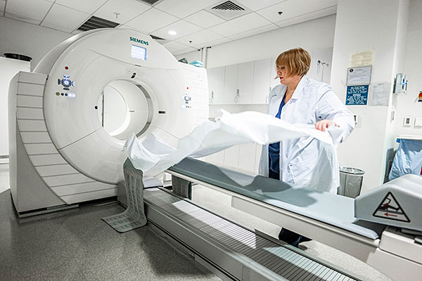
CT scan — A CT scan uses X-rays to create detailed cross-section images of your bones, organs and tissues. It’s quick, painless and can help providers pinpoint injuries, infections or internal conditions.
MRI — MRI uses powerful magnets (not radiation) to create high-resolution images, especially of soft tissues like the brain, spine, joints and muscles. Our open-bore 3.0 Tesla MRI allows for a more comfortable experience — ideal for people who are claustrophobic or have a larger body size — without compromising image quality.
PET scan — PET scans show how your cells are functioning. This safe and noninvasive test helps detect early signs of cancer, neurological conditions and heart disease by highlighting abnormal activity in the body.
Nuclear medicine imaging — These scans use a small amount of radioactive tracer to see how your organs and tissues are working. Despite the name, these scans are very safe and are often the least invasive way to diagnose or keep track of a condition.
Whole body MRI — This scan provides a head-to-thigh view of your health. It may detect early signs of cancer, inflammation or other changes — even before symptoms appear. It’s a good option if you want a proactive check-in with your body because there’s no radiation exposure. Scans include:
- Head: brain, skull, sinuses, eyes, ears
- Neck: major arteries, upper airway, lymph nodes, glands
- Chest: lungs (using low-dose CT), lymph nodes, heart, large blood vessels
- Abdomen: kidneys, liver, spleen, adrenal glands, gallbladder, pancreas
- Pelvis: uterus, ovaries, prostate
- Spine: spinal cord, vertebrae, discs
Coming soon — Cardiac and calcium scoring to assess heart health.
