



A bubble study is an ultrasound test that uses a contrast agent, called agitated saline, to examine blood flow in your heart and lungs. The saline contains tiny bubbles (microbubbles) that appear in the images and can show if blood is flowing the wrong way. It’s a common test for diagnosing a patent foramen ovale, which is a hole in your heart.
Advertisement
Cleveland Clinic is a non-profit academic medical center. Advertising on our site helps support our mission. We do not endorse non-Cleveland Clinic products or services. Policy
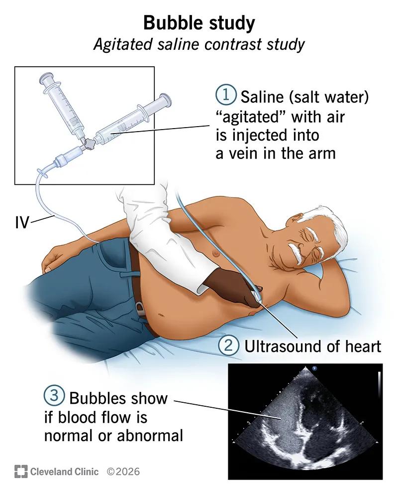
Image content: This image is available to view online.
View image online (https://assets.clevelandclinic.org/transform/51ced08e-ffea-4172-ad7f-8bbe713994d3/Bubble-Study-Illustration)
A bubble study, also called a bubble echocardiogram or agitated saline contrast study, is a type of ultrasound test that checks blood flow through your heart and lungs. It can show if blood is flowing through an abnormal passage called a shunt. The shunt might connect the right and left sides of your heart. Or it might link your pulmonary arteries and veins, which carry blood between your heart and lungs.
Advertisement
Cleveland Clinic is a non-profit academic medical center. Advertising on our site helps support our mission. We do not endorse non-Cleveland Clinic products or services. Policy
A bubble study can be done along with any of these imaging tests:
You may need a bubble study if you have signs or symptoms of a shunt, like low blood oxygen levels and shortness of breath, or you’ve had a stroke without a clear cause.
Blood should normally pass through your lungs to pick up oxygen and get rid of waste. In addition, capillaries in your lungs filter out tiny blood clots so they don’t flow back through your body and cause problems. A bubble study can show if any blood skips this step. The results guide treatment.
This test most commonly diagnoses:
Advertisement
A bubble study is similar to an echocardiogram without contrast. You lie on your side, and a healthcare provider takes ultrasound images of your heart. But there’s a key difference. During a bubble study, your provider injects a substance called agitated saline into a vein in your arm. It acts as a contrast agent. That means it enhances the ultrasound images to better show blood flow.
Saline is salt water. Your provider “agitates” it by quickly mixing it with a small amount of air. This makes tiny bubbles, called microbubbles. Your provider may also add a bit of your own blood to help make more bubbles.
The bubbles flow through your veins and into your heart. Your provider looks at exactly where they appear on the ultrasound images. This shows them if blood is flowing in normal or abnormal ways.
You usually don’t need any special prep for a transthoracic echo or transcranial Doppler ultrasound. For a transesophageal echo, you’ll need to fast (typically for at least six hours) beforehand. Your provider may also give you other instructions.
The exact steps can vary based on the type of imaging test you’re getting. To perform a bubble study with a transthoracic echo, your healthcare provider will:
You can usually go back to your usual activities right after the test and drive yourself home. But if you have a transesophageal echo, the process is a little different. Because you’re sedated, you’ll have to stay for a bit after the test so your provider can monitor you. And you can’t drive for 24 hours. So, you’ll need someone to drive you home.
Advertisement
Your healthcare provider will schedule a time with you to go over the test results.
A bubble study is a safe and low-risk test. A very rare complication during the test is a stroke, which is typically mild and treatable.
It’s possible for a bubble study to produce false negatives. That means it shows there’s no shunt, but there actually is one. This is more likely to happen if you have raised pressure in the right side of your heart, due to a condition like mitral valve disease. Your healthcare provider will consider your medical history when interpreting the test results.
The results of a bubble study can be either positive or negative. Here’s what those mean:
Providers look closely at the echo images to learn more about the exact type of shunt you have. For example:
Advertisement
The larger the shunt, the more bubbles will be visible in the images.
You may need treatment for a shunt. Your healthcare provider may recommend:
Your provider will explain any next steps and what you can expect.
Bubbles aren’t just for play. In the medical world, special “microbubbles” in agitated saline can help your healthcare provider see how blood flows through your heart and lungs. A bubble study offers valuable — even lifesaving — answers to your symptoms. Don’t hesitate to ask your provider any questions you have before getting one done.
Advertisement



Sign up for our Health Essentials emails for expert guidance on nutrition, fitness, sleep, skin care and more.
Learn more about the Health Library and our editorial process.
Cleveland Clinic’s health articles are based on evidence-backed information and review by medical professionals to ensure accuracy, reliability and up-to-date clinical standards.
Cleveland Clinic’s health articles are based on evidence-backed information and review by medical professionals to ensure accuracy, reliability and up-to-date clinical standards.
Breathing issues can affect your life in many ways. Cleveland Clinic’s respiratory therapy program treats your symptoms so you can feel better day to day.
