A foot X-ray is a test that produces an image of the anatomy of your foot. Your healthcare provider may use foot X-rays to diagnose and treat health conditions in your foot or feet. Foot X-rays are quick, easy and painless procedures. A radiologic technologist will place your leg on an X-ray table and then take multiple pictures of it.
Advertisement
Cleveland Clinic is a non-profit academic medical center. Advertising on our site helps support our mission. We do not endorse non-Cleveland Clinic products or services. Policy
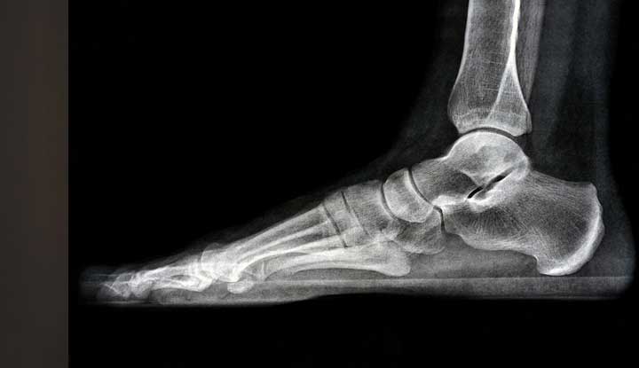
Image content: This image is available to view online.
View image online (https://my.clevelandclinic.org/-/scassets/images/org/health/articles/23500-foot-x-ray)
A foot X-ray is a test that creates a black-and-white picture of the inside of your foot. The image displays the soft tissues and bones of your foot. These bones include your ankle bones (tarsal bones), the front end of your foot (metatarsal bones) and your toes (phalanges). A foot X-ray is also called a foot series or foot radiograph. Healthcare providers use foot X-rays to diagnose medical conditions in your ankles or feet.
Advertisement
Cleveland Clinic is a non-profit academic medical center. Advertising on our site helps support our mission. We do not endorse non-Cleveland Clinic products or services. Policy
X-rays use a form of radiation called electromagnetic waves. These waves create a picture of the inner structure (anatomy) of your body. X-rays were first discovered in 1895, making them the oldest type of medical imaging used. The first X-ray took a picture of human tissue in 1896. X-rays are also the most frequently used type of imaging.
A foot X-ray may be used to look for signs of possible injury to your foot or ankle, especially after an accident. Your healthcare provider will use a foot X-ray to diagnose possible health conditions in your feet or ankles. These conditions may include:
If you have unexplained swelling, pain or tenderness in your foot or ankle, a foot X-ray can help your healthcare provider diagnose the cause.
Foot X-rays are often used after a broken ankle bone has been set. The X-ray will show your healthcare provider if the bone was set properly. Bones need to be set in the proper alignment to heal correctly.
A radiologic technologist, or X-ray technician, will perform your foot X-ray. These specially trained healthcare providers have education in radiation protection, patient care, radiation exposure, radiographic positioning and radiographic procedures.
Advertisement
X-rays send small beams of radiation through your body. The X-rays create a picture on special photographic film or a digital platform.
Your body parts vary in thickness, so they absorb different degrees of radiation. Calcium in your bones absorbs more radiation, so your bones look white on X-rays. Your soft tissues, including your organs, fat and muscles, absorb less radiation. So these tissues look different shades of gray. Air looks black on X-rays.
Foot X-rays don’t require much preparation. Before the test, you’ll have to remove any clothing, shoes, jewelry or metal objects that may get in the way of clear X-ray images.
It’s also important to tell your radiologic technologist if you’re pregnant or if you could be pregnant. There’s a chance your growing baby (fetus) could be exposed to radiation. Your healthcare provider will decide if you need the X-ray. If the X-ray must be done because of an urgent need, precautions will be taken to minimize radiation exposure to your baby.
Before your X-ray, the technologist will explain the process to you. They will answer any questions you have and make sure you’re ready before starting the procedure.
A radiologic technologist will perform your foot X-ray in an X-ray room. This may be in your healthcare provider’s office or a hospital radiology department. Once in the X-ray room, the technologist may place a lead apron on your lap to protect your reproductive organs from radiation exposure. This is especially important if you’re pregnant or could be pregnant. The X-ray room may be a little chilly, but the entire procedure should take less than 15 minutes.
The technologist will place your leg on the X-ray table. Then the technologist may put positioning equipment such as sandbags or pillows around your leg or foot to keep it from moving. It’s important to keep very still during the X-ray because movement can make the X-ray images blurry.
Your technologist will place an X-ray film holder or digital recording plate under the X-ray table. Then they will go behind a wall or into a special room to activate the X-ray machine. Your technologist may reposition your foot several times to get images from various angles. Three separate images are usually taken to make sure they get all views. The technologist will take one image from the side, one image from the front and one image at a 45-degree angle between the front and side views. Let your technologist know if you’re experiencing any pain. They will assist you and make you as comfortable as possible throughout the test.
After your foot X-ray, your radiologic technologist will probably ask you to wait a couple of minutes while they check the images. They want to make sure the images aren’t blurry before sending you on your way. If any images are blurry, they will retake them immediately.
A doctor called a radiologist will then review the images. Radiologists are trained to study X-ray images and figure out what they mean. Once the radiologist has reviewed the results, they will send them to your healthcare provider. Your healthcare provider will talk with you about the results and determine the correct treatment for your condition. You may be referred to a foot and ankle surgeon, who has specialized training in foot and ankle conditions.
Advertisement
If your healthcare provider or specialist wants to see additional views of your foot or ankle, you may have to return for a follow-up X-ray. You may also have to return to monitor your condition and keep track of any changes that occur over time.
X-rays provide a quick and simple way for your healthcare provider to diagnose possible health conditions in your foot or feet. There’s only a slight amount of radiation exposure in a foot X-ray, and the radiation passes right through you. In addition, X-rays rarely cause side effects.
Women who are pregnant have a somewhat elevated risk of issues with radiation exposure. You should always tell your radiologic technologist if you’re pregnant or think you might be pregnant. You may wear a lead apron to protect your reproductive organs from radiation exposure. Children have a somewhat higher risk as well. Lower amounts of radiation can be used on children.
Excessive exposure to radiation carries a minor risk of cancer. However, healthcare providers agree the benefit of an accurate diagnosis outweighs the risk of radiation exposure. If you’re concerned about the amount of radiation you’ll be exposed to, ask your technologist.
The results of X-rays taken during emergencies are often available immediately. For non-emergencies, your radiologist will typically have your results to your healthcare provider within one to two days. Then your healthcare provider will discuss the results with you and go over treatment options.
Advertisement
Pain, swelling and tenderness in your foot or ankle can knock you off your feet and disrupt your life. Your healthcare provider may recommend a foot X-ray if they need help determining what’s going on. Once your healthcare provider gets results back from a radiologist, they can diagnose your condition. They will speak with you about possible treatment options, and hopefully, you’ll be back on your feet before you know it.
Advertisement


Sign up for our Health Essentials emails for expert guidance on nutrition, fitness, sleep, skin care and more.
Learn more about the Health Library and our editorial process.
Cleveland Clinic’s health articles are based on evidence-backed information and review by medical professionals to ensure accuracy, reliability and up-to-date clinical standards.
Cleveland Clinic’s health articles are based on evidence-backed information and review by medical professionals to ensure accuracy, reliability and up-to-date clinical standards.
When you need a clear picture of what’s happening inside your body, the Cleveland Clinic imaging team is here for you.
