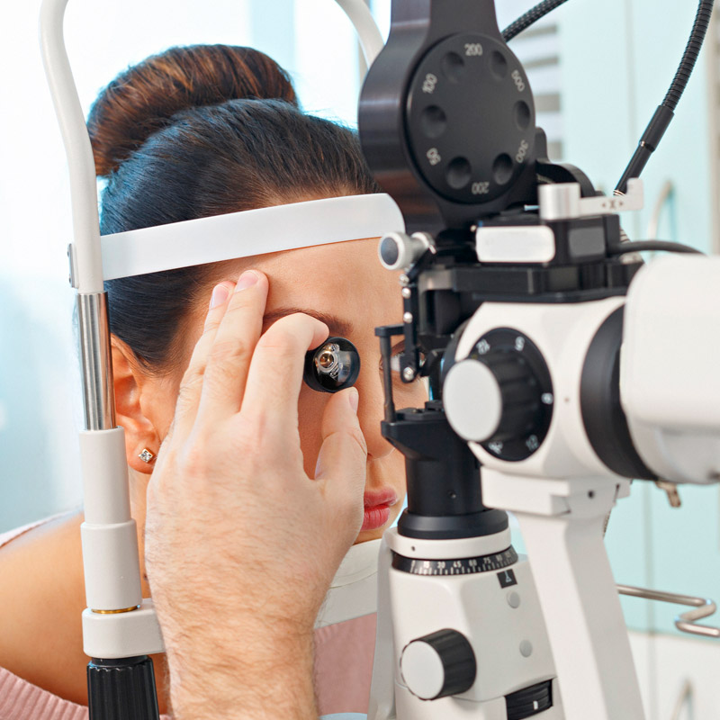
Gonioscopy is an eye test that checks for signs of glaucoma. An eye specialist does this using a special lens and slit lamp to look at your eye’s drainage angle (anterior chamber angle). If the drainage angle is too narrow or closed, you may have glaucoma.
Advertisement
Cleveland Clinic is a non-profit academic medical center. Advertising on our site helps support our mission. We do not endorse non-Cleveland Clinic products or services. Policy

Image content: This image is available to view online.
View image online (https://my.clevelandclinic.org/-/scassets/images/org/health/articles/gonioscopy)
Gonioscopy is a noninvasive diagnostic test that eye care specialists, especially ophthalmologists, can use to check for several conditions, like glaucoma. To do this test, your eye care specialist will use special tools to look inside your eyes. This test is part of how they make sure your eye’s drainage system works properly.
Advertisement
Cleveland Clinic is a non-profit academic medical center. Advertising on our site helps support our mission. We do not endorse non-Cleveland Clinic products or services. Policy
There are two correct ways to pronounce “gonioscopy:”
Your eye care specialist can use glaucoma tests, including gonioscopy, to diagnose or rule out certain types of glaucoma or conditions that can cause glaucoma-like effects. That includes:
Glaucoma happens when you have high pressure inside your eye (intraocular pressure). If the pressure is too high or stays high for too long, it can cause permanent, severe eye damage and vision loss.
Gonioscopy lets your eye care specialist look directly inside the anterior chamber, a fluid-filled space behind your cornea and in front of your iris. Your specialist uses gonioscopy to look for the drainage angle, a ring-shaped area where your iris and sclera (the white of your eye) meet. The angle needs to be wide enough for aqueous humor fluid inside the anterior chamber to drain.
Gonioscopy uses a combination of mirrors and/or magnifying lenses to let your eye care specialist see around the edge of the sclera and into the drainage angle. It’s like aiming a mirror so the reflection shows you what’s around a corner.
Advertisement
There are two types of gonioscopy lenses:
Gonioscopy is usually a test that your eye care specialist can do anytime. It typically doesn’t require any preparation on your part. One exception is if you wear contacts. If you do, ask your eye specialist if you need to stop wearing them within a certain timeframe before your gonioscopy.
If you need gonioscopy as part of a surgery, your eye care specialist will give you specific instructions on how to prepare.
What you can expect during gonioscopy depends on which type you’re undergoing.
Direct gonioscopy usually only happens in hospitals, and it’s common for you to receive general anesthesia before these procedures start. That means you’ll be asleep while direct gonioscopy and related procedures happen.
Indirect gonioscopy is most likely to happen in an eye care specialist’s office, and it has several similarities to a slit lamp exam. Those exams involve you sitting in a chair facing your specialist.
Before they start a gonioscopy exam, your eye care specialist will apply numbing drops or similar medications to the surface of your eye. Your corneas are extremely sensitive to anything touching them, so numbing drops help you feel more comfortable and not want to blink during the exam.
After numbing your eye, your eye specialist will use wetting drops or similar substances on the surface of the lens that will touch your eye. The wetting substances push air out from under the lens, preventing air bubbles from interfering with what your specialist can see. They also help the lens move smoothly on the surface of your eye when your specialist needs to adjust its position.
Once the lens is ready, your eye specialist will touch the lens to your eye and hold it in place with one hand. They’ll then use the other hand to operate the slit lamp device. The device has a bright light that lights up the inside of your eye while they use a magnifier to look closely inside your eye. Your specialist will tell you to look straight ahead or in a specific direction. They may also use a technique called “dynamic gonioscopy,” where they use the lens to gently press on your eye surface. That light pressure lets them check if your iris stays stuck against your cornea or lens (synechiae).
Advertisement
Once your provider finishes looking around the entire outer edge of your iris, they’ll remove the lens from your eye. They may also rinse your eyes with wetting drops or similar substances. It’s common for your eyes to tear up or water when this happens. Your specialist will give you a tissue or something similar to help you dry any tears or wipe away excess solution on your face.
Gonioscopy may or may not involve medications that dilate your eyes, and your provider may want to look inside your eyes before and after using dilating medications. Your specialist can tell you more about whether they think dilating your eyes is necessary.
Gonioscopy itself doesn’t have any side effects or risks. The medications they use to numb and dilate your eyes may have some side effects. Your eye care specialist can tell you more about any possible side effects from those medications.
If you’re awake during gonioscopy, your eye(s) should be numb. That means you won’t feel pain or discomfort during this exam. You may feel slight pressure similar to what it feels like if you rub your eyes during certain parts of the exam.
If you undergo direct gonioscopy during a surgery or similar procedure, your eye care specialist will talk to you after you wake up. They’ll explain any results from the procedure and other details you should know.
Advertisement
If you undergo indirect gonioscopy in your eye specialist’s office, they can tell you what they see during or immediately after they finish your exam. They can tell you more about what they saw and what that means. They can also give you recommendations on treatment options — if necessary — and give you guidance on anything you may need to do afterward.
If your eye specialist sees a narrowed drainage angle or any other evidence that the aqueous humor isn’t draining properly, they’ll talk to you about treatment options.
Treating glaucoma is important because the pressure buildup can cause permanent damage to the optic nerve in the back of your eye. That can cause permanent vision loss.
If your provider sees that your eye’s drainage angle is closed, that’s called angle-closure glaucoma. This condition is a medical emergency because it can cause sudden vision loss (which can quickly become permanent). Your eye care specialist will recommend emergency treatment and help you get to a medical facility that can treat this condition.
Glaucoma is one of the leading causes of vision loss worldwide. About 90% of glaucoma cases are chronic, meaning they develop gradually. That’s one reason you should get regular eye exams every one to two years (or at least annually if you’re over 40). You may also need more frequent eye exams, including gonioscopy, if you have any of the following glaucoma-related conditions or circumstances:
Advertisement
You should call your provider if you notice glaucoma symptoms. Symptoms that mean you should call your eye specialist include:
Some glaucoma symptoms mean you need immediate medical attention. Those symptoms include:
Gonioscopy is a valuable exam, but it isn’t enough to detect or diagnose glaucoma. There are many factors that go into determining glaucoma, and gonioscopy just provides one clue to the puzzle.
Indirect gonioscopy should take five minutes or less. But your appointment will likely last longer, especially if you need other tests to confirm or rule out any issues.
The time it takes to do direct gonioscopy can vary greatly depending on the procedure it happens with. Your eye care specialist can tell you more about how long this procedure should take.
Gonioscopy may have a complicated name, but the test itself is simple. It’s a noninvasive, painless way for your eye care specialist to look for signs of conditions that could cause permanent vision loss. This test is usually something your eye specialist can do in their office, and they can tell you the results immediately.
If you’re worried about any part of how this exam will go, tell your eye care specialist. They can explain the test, help you feel less anxious about it and help you understand what they saw during the exam. This test can make a big difference and protect your eyes from potentially damaging conditions.
Learn more about the Health Library and our editorial process.
Cleveland Clinic's health articles are based on evidence-backed information and review by medical professionals to ensure accuracy, reliability, and up-to-date clinical standards.
Cleveland Clinic's health articles are based on evidence-backed information and review by medical professionals to ensure accuracy, reliability, and up-to-date clinical standards.
Getting an annual eye exam at Cleveland Clinic can help you catch vision problems early and keep your eyes healthy for years to come.
