Cardiac Sarcoidosis - Part 2: Diagnosis and Treatment

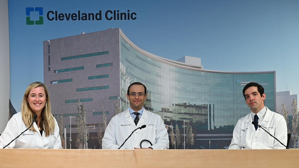
Welcome to Part 2 of the 2-part series for cardiac sarcoidosis. In Part 1, Dr. Christine Jellis, Dr. Manuel Ribeiro Neto and Dr. Ziad Taimeh discussed a team approach to caring for patients with cardiac sarcoidosis. In Part 2, learn about diagnosis, testing and treatments for cardiac sarcoidosis.
Learn more about the Sarcoidosis Center
Subscribe: Apple Podcasts | Buzzsprout | Spotify
Cardiac Sarcoidosis - Part 2: Diagnosis and Treatment
Podcast Transcript
Announcer:
Welcome to Love Your Heart, brought to you by Cleveland Clinic’s, Sydell and Arnold Miller Family, Heart Vascular & Thoracic Institute. These podcasts will help you learn more about your heart thoracic and vascular systems, ways to stay healthy and information about diseases and treatment options. Enjoy.
Christine Jellis, MD, PhD:
Thank you for joining us to the Heart, Vascular and Thoracic Institute of Cleveland Clinic. I'm pleased to welcome you here today to talk about cardiac sarcoidosis. I'm Christine Jellis, I'm one of the staff cardiologists here in the Section of Cardiac Imaging. And I am fortunate to have with me two real experts in cardiac sarcoidosis, Dr. Manny Ribeiro, Dr. Ziad Taimeh. Manny is a pulmonologist and the Director of our Sarcoid Center, and Ziad is an expert in heart failure. And together we are three members of our multidisciplinary cardiac sarcoidosis team, of which there are many others. And hopefully we can share with you some of the insights that we've learned along the way today.
Christine Jellis, MD, PhD:
Firstly, we'd like to focus on diagnosis of cardiac sarcoidosis because, I think often patients present with symptoms, but we also see patients who have a history of pulmonary sarcoidosis, or sarcoidosis elsewhere in the body, who are concerned that they may have or may develop cardiac sarcoidosis. So I'd love to share with you some of our insights on what we've learned and the algorithm that we work through, to try and figure out with a degree of certainty, of course, whether people have cardiac sarcoidosis. So guys, if I were to come into your office and I have a history of pulmonary sarcoidosis, and I don't have any cardiac symptoms, but I'm worried about I might get cardiac sarcoidosis. What advice would you be giving me?
Manuel Lessa Ribeiro Neto, MD:
Chris, I think starting with the easiest things, right? So I think one thing that everybody agrees, experts and different guidelines is that some type of screening needs to happen for cardiac sarcoidosis. What tests specifically we do in that screening, that varies a little bit, but we should definitely screen for cardiac sarcoidosis in patients with those symptoms. At least one EKG, and this has been our approach here, I think that needs to be done. But depending on how many symptoms the patients have, we should expand that workup. For example, if patients are having palpitations, feeling the heart beating too fast or too slow, something that is a little bit more than expected, then I think it's reasonable to expand that work up and get a Holter monitor or sometimes even an echocardiogram. But I think the safest thing to say is that definitely screening should be done with at least one EKG.
Christine Jellis, MD, PhD:
Perfect. I would definitely agree. Ziad if we are suspicious, then on the EKG or from symptoms that someone has got cardiac sarcoidosis, what would be your next step in determining that further?
Ziad Taimeh, MD:
I think it would be very important for us to get a comprehensive cardiac history to understand, are there any other heart conditions that might be confounding the picture and be the cause of these signs and symptoms. After that, we would need to get a detailed imaging about, looking at the heart muscle itself. Like my colleague said about an echocardiogram or maybe a specialized echo with strain analysis to see if there are any little bit more in depth abnormalities. And if all is, well, then we would need something little bit more specific looking at the heart muscle itself. We like to start with a cardiac MRI and I refer to you to give us a little bit more feedback, but I always feel that the cardiac MRI would be the next best step to tell us whether we have any heart involvement with at least scarring or inflammation.
Christine Jellis, MD, PhD:
I would definitely agree, I think. I'm obviously an imaging cardiologist, so I have to declare my bias up front. But I think an echo is a really easy test. It's a portable test, it's readily available, there is no radiation. Most people can access that locally, as well as in specialized centers like ours. And we get a lot of good information about the pumping function of the heart, and we often use the term ejection fraction to describe the amount of blood that's ejected out of the heart with every beat. And usually the convention is accepted that anything over 55% is classed as normal. So if someone has an ejection fraction of 55% or more, a normal looking heart and nothing to suggest that there's anything else going on, like coronary artery disease or valvular heart disease, then in many cases we could sort of leave things alone.
Christine Jellis, MD, PhD:
But if we then find that there are abnormalities and there is a depressed ejection fraction, or there is another regional wall motion abnormality on the echo, then that starts to give us a few little red flags that perhaps this person does have cardiac sarcoidosis and warrants further evaluation. Now sometimes on the EKG or the Holter monitor, we see conduction abnormalities that perhaps make us more likely to order further imaging tests in addition to the echo. But I think doing it in that step wise fashion is, is generally the way that most of us operate. I'm obviously an MRI lover, so I think it has a really useful role, it can help us determine not only function of the heart, but look at the tissue characteristics of the muscle to see if there's any active inflammation or scar. And that I hope that you guys find useful in determining treatment for these patients.
Ziad Taimeh, MD:
Absolutely. I mean, at the end of the day, we have to build a story. We have to build a case for cardiac sarcoidosis diagnosis. Because like we said earlier, it's a great masquerader, so there are other causes that can have similar presentation. So starting with the screening, going with the actual medical examination, going with EKGs Holter monitor for arrhythmias, then MRI. And then if there are abnormalities in the MRI, we try to build a case of inflammation using cardiac PET FDG scans. And at the end of the day tissue diagnosis I think is very, very important, and we collaborate with our pulmonary colleagues to see if we can't take tissue sample of the lymph nodes in the chest or anywhere that's accessible.
Ziad Taimeh, MD:
And in most I want to say extreme cases but at the end of the day, we could as access the heart with a heart biopsy, which is safe to do, we do it here on the right side or on the left side and collaborations with our electrophysiology colleagues to see if we can take a sample from those areas that are lighting up on the cardiac MRI or the cardiac PET scans.
Christine Jellis, MD, PhD:
Couple of things you mentioned there. I think that was a great overview, because I think tissue is always helpful. If we can confirm a diagnosis with a biopsy of a lymph node, which is often the most readily available, or the myocardium in some circumstances, then that can solidify our diagnosis and its obviously integral into making sure we treat the patient appropriately. Do you guys want to give your perspectives on the different types of biopsies because patients may be offered endo myocardial biopsies or lymph node biopsies. Manny, you do the endo bronchial biopsies. Could you tell us a little bit about that and the advantages to that over perhaps a cardiac biopsy, although a cardiac biopsy in experienced hands like Ziad’s is a valuable tool.
Manuel Lessa Ribeiro Neto, MD:
Correct, no, I completely agree, Chris. I think a few important points, one is, anytime that it's possible, we should try to get a biopsy. We should try to confirm under the microscope that we are dealing with sarcoidosis, there are many diseases that can mimic sarcoidosis. So it's very important to try to do the first step. So that's one important message. The other important decision point is, do we have an easy place to biopsy outside of the heart or don't, or maybe the heart is really the only place to target. If we have an easy place to biopsy outside of the heart, and usually a PET scan, for example is a good imaging test to guide us. So if we have that easy place outside of the heart, we should target that first, as you mentioned, a very common location outside of the heart that we can do biopsies to make the diagnosis of sarcoidosis are the lymph glands in the chest or the lungs.
Manuel Lessa Ribeiro Neto, MD:
So if we see any abnormality in the PET scan in one of those areas, lymph glands in the chest or the lungs, we recommend a bronchoscopy. So this is an endoscopy that we do in our bronchoscopy suite under general anesthesia usually, and we do small biopsies of those areas. So this is kind of the easier scenario when we can see easy places to biopsy outside of the heart. But the other scenario is when there's nothing obvious to biopsy outside of the heart. In those cases, most of the time, if we think a biopsy is needed and a lot of times we do, then we go to an endo myocardial biopsy.
Manuel Lessa Ribeiro Neto, MD:
But one thing that we have been doing here recently is, we've been successful in targeting very small lymph nodes in the chest. Lymph nodes that look normal on the PET scan or on the CT scan that maybe in other places, we wouldn't think of biopsying those lymph nodes. But here in our experience, we can find granulomas, we can find sarcoidosis in about 30% of those patients. So if we see small lymph glands, five, six, seven millimeters, even if they look normal on imaging, I think that bronchoscopy is a good first step. If they are normal or if we don't think that bronchoscopy is a good idea, then I refer to my friend Ziad to do the heart biopsy.
Christine Jellis, MD, PhD:
Ziad, do you mind telling us a little bit about that process? And then I'd like to just circle back talking about dietary preparation for the PET at the end, because I think that's something really important that we all rely on.
Ziad Taimeh, MD:
Yeah, absolutely. So endo myocardial biopsy or heart biopsy, it's basically a 30 minute procedure to be able to access the heart and get very small, tiny sub millimeter tissue samples that we can put under the microscope. It is fairly straightforward to do it from the right side of the heart. It goes through the vein access through the neck. One of the challenges, sarcoidosis attacks the heart in a patchy fashion. So to be able to get the catheter or the bioptome to reach that area is quite difficult. So the yield of having a positive biopsy through these random right side biopsies, is less than 10%. That's why we generally don't do that unless we feel comfortable that the actual inflammation really is attacking an area where we can tackle with that biopsy.
Ziad Taimeh, MD:
In other cases we have done in the past is we go with the left side biopsy, the left side of the heart. In collaboration with our electrophysiology colleagues, we gain access to the left ventricle. We look at the MRI and look at the cardiac path and do the voltage mapping with their assistance. And once we do that, we'll be able to find out what areas in the ventricle are abnormal for us to sample, and that would increase the yield from less than 10% to almost up to 70% of the cases. So it has a better diagnostic yield. It's a little bit more challenging because it involves a multidisciplinary team to actually get the biopsy itself. But in certain cases it is important to actually get.
Christine Jellis, MD, PhD:
I'm really pleased that you mentioned the EP team and I'd also like to acknowledge our Nuclear Cardiology team, because they've been really integral to our success as a multidisciplinary team. And obviously there are circumstances where patients with cardiac sarcoidosis develop heart block may need a pacemaker, have ventricular arrhythmias or depressed ejection fraction and may benefit from a defibrillator. And these are all things that we discuss in our regular team meetings, because we want to make sure that we're availing our patients of all these appropriate therapies.
Christine Jellis, MD, PhD:
Lastly, it would be remiss of me not to mention dietary preparation for PET scans, because I think increasingly we are wanting to follow patients up on a regular basis to make sure that they're getting an adequate response to treatment and to determine exactly how long that treatment should be. And I think echo is always a good workhorse in junction with an EKG obviously, but I think PET imaging has really revolutionized the way that we follow up a lot of these patients looking for residual inflammation and one of the keys to this is doing a scan in a center that has great experience and technical expertise in doing those scans. And then making sure that you, as the patients are adequetly prepared for the scans because you don't want to go to all the effort of coming for the scan, the expense of and the travel and all of that, only to be not be adequately prepared from a dietary perspective.
Christine Jellis, MD, PhD:
So guys, let's just in the last couple of minutes, just wanted to really plug that the preparation is key because if we can get accurate information from those tests, then it really pulls everything together.
Manuel Lessa Ribeiro Neto, MD:
Yeah. I think it's a great point, Chris. And to make it simple, I think the most important message is no sugar, right? No sugar, no carbohydrates for at least 24 hours before the test, one day before the test. After midnight of that day of the test then nothing per mouth, right. But it is important to remember that on the day prior, no sugar, no carbohydrates. Usually when we schedule that test here, our patients will get a list of all of the specific food items or meals that are allowed and are not allowed, but to make it simple, anything with sugar or carbohydrates in it, just don't don't get it, no potatoes, no pasta, no soda, right. And the reason for that is going back a little bit to that granuloma.
Manuel Lessa Ribeiro Neto, MD:
So the PET scan that we do, we give a specific type of sugar, a specific type of glucose. And we try to detect that inflammation. We try to make those granulomas take or use the glucose. And then we can detect that in the test. The problem is, the heart muscle uses a lot of sugar, a lot of glucose as well. So we need to do that diet to suppress the heart utilization of the sugar. So only the sarcoidosis is only the granuloma is using that sugar and being detected on that PET scan. So no sugar, I think is good advice in general.
Christine Jellis, MD, PhD:
Definitely. And Ziad, just finally for our patients who do have evidence of significant cardiac dysfunction, I think we mentioned device therapy. And I just want to make sure that we put that out here as something that should be considered in patients with depressed ejection fractions, and just have you comment on, do we do that any differently in the sarcoidosis population compared to the general population?
Ziad Taimeh, MD:
Absolutely. I think it's very important to understand is that it doesn't matter what the ejection fraction is. A patient with sarcoidosis remain at risk of arrhythmias and not only arrhythmias, but heart block where the sarcoidosis cells attack the conduction system of the heart in needing a pacemaker, not just a defibrillator. So the consensus in the medical community now is to consider a dual chamber defibrillator in all patients with diagnosis of cardiac sarcoidosis regardless to the ejection fraction. Now it's very critical if the ejection fraction drops below 35%, but the medical guidelines support to consider in any patient with sarcoidosis. Here at the Cleveland Clinic, we have been a little bit more into doing dual chamber defibrillators in most patients with sarcoidosis, regardless of the ejection fraction.
Ziad Taimeh, MD:
Now in certain cases where patients is very young or they have issues with getting a defibrillator, then further prognostication is very important and acknowledging our electrophysiology colleagues by doing Holter monitors, Zio patching for a couple weeks to look at arrhythmias. And in certain cases do an EP study, try to induce those arrhythmias in the cath lab, and see if those are patients at risk, I think is very reasonable.
Christine Jellis, MD, PhD:
Thank you. Well, I hope that this has given you some insights about how we think approaching cardiac sarcoidosis. As you can probably tell, we enjoy working together as part of a multidisciplinary team. And we look forward to bringing you updates on our team and the work that we are doing, including the research in that area. Please let us know if we can be of any assistance to you or your loved ones with sarcoidosis.
Announcer:
Thank you for listening. We hope you enjoyed the podcast. We welcome your comments and feedback. Please contact us at heart@ccf.org. Like what you heard, subscribe wherever you get your podcasts or listen at clevelandclinic.org/loveyourheartpodcast.

Love Your Heart
A Cleveland Clinic podcast to help you learn more about heart and vascular disease and conditions affecting your chest. We explore prevention, diagnostic tests, medical and surgical treatments, new innovations and more.


