A pelvis X-ray is a test that produces an image of the anatomy of your pelvis. Your healthcare provider may use pelvis X-rays to diagnose and treat health conditions involving your pelvis. Pelvis X-rays are quick, easy and painless procedures.
Advertisement
Cleveland Clinic is a non-profit academic medical center. Advertising on our site helps support our mission. We do not endorse non-Cleveland Clinic products or services. Policy
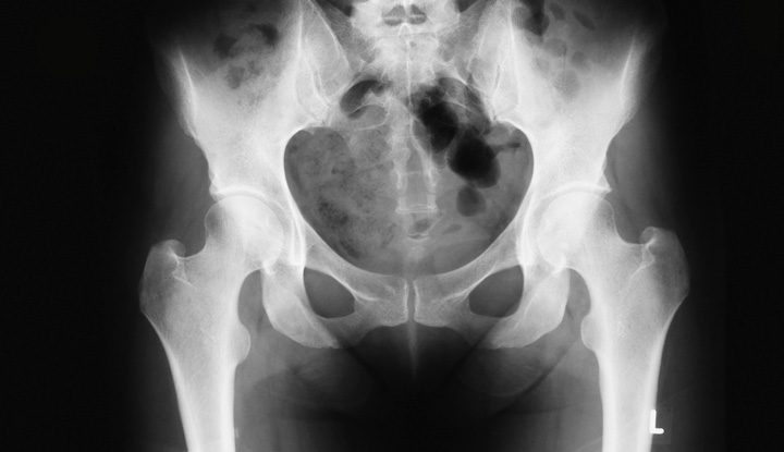
Image content: This image is available to view online.
View image online (https://my.clevelandclinic.org/-/scassets/images/org/health/articles/23519-pelvis-x-ray)
A pelvis X-ray (radiograph) is a medical imaging test that creates a black-and-white picture of your pelvic bones. Your pelvic bones include your hip bones (ilium, ischium and pubis), the triangle-shaped bone at the base of your spine (sacrum) and your tailbone (coccyx). Healthcare providers use pelvis X-rays to diagnose and treat medical conditions that affect the area in and around your pelvis.
Advertisement
Cleveland Clinic is a non-profit academic medical center. Advertising on our site helps support our mission. We do not endorse non-Cleveland Clinic products or services. Policy
X-rays use a form of radiation called electromagnetic waves. These waves create a picture of the inner structure (anatomy) of your body. X-rays were first discovered in 1895, and the first X-ray took a picture of human tissue in 1896. X-rays are the most commonly used type of medical imaging.
Healthcare providers can use pelvis X-rays to find the possible cause of swelling, pain or deformity in your pelvis, hips or upper leg areas. They can use pelvis X-rays to diagnose health and medical conditions involving your pelvis. These conditions may include:
In addition, if you need pelvic surgery, your healthcare provider will want X-rays before the procedure. They’ll also want you to have routine follow-up X-rays afterward to track your condition.
A radiologic technologist, also known as an X-ray technician, will perform your pelvis X-ray. Radiologic technologists are healthcare providers specially trained in patient care, radiation exposure, radiation protection, radiographic positioning and radiographic procedures.
Advertisement
X-rays send small beams of radiation through your body. These beams of radiation produce a picture on special photographic film or a digital platform.
Your body parts vary in thickness, so they take in different amounts of radiation. Calcium in your bones takes in more radiation, so your bones appear white on the X-ray. Soft tissues take in less radiation, so they appear in different shades of gray. Air appears black. If you have a broken bone, the bone will appear white and a black line running through the bone will show the fracture.
Pelvis X-rays don’t require too much preparation. You should wear comfortable clothing that doesn’t contain any metal. You’ll be asked to remove any clothing, jewelry, belts or other objects containing metal. Metal can interfere with getting clear X-rays.
If you’re pregnant, it’s very important to tell your radiologic technologist. Pelvis X-rays use a very small amount of radiation, but there’s a chance your growing baby (fetus) could be exposed to it. Your healthcare provider will decide if you need the X-ray. If the X-ray is urgent or necessary, precautions will be taken to minimize radiation exposure to your baby.
Before your X-ray, your radiologic technologist will explain how the X-ray will work. If you have any questions about the process, your technologist can answer them for you.
Your pelvis X-ray will be performed by a radiologic technologist in your healthcare provider’s office or a hospital radiology department. The X-ray room will contain a table with an X-ray machine hanging from the ceiling or wall. Once in the X-ray room, your technologist may give you a lead apron to wear to protect you from radiation exposure. The procedure may take 10 minutes or more.
Your technologist will place a digital recording plate under the X-ray table. They’ll have you lie down on your back on the table. You’ll need to keep very still during the procedure. Any movement can cause the X-ray images to show up blurry. The technologist may ask you to hold your breath while they’re taking the images.
Your technologist will go into a small room or behind a wall to control the X-ray machine. They’ll return to reposition you for additional images. A normal pelvis X-ray includes at least one or two different images. Your technologist will take one image with your legs straight (anteroposterior view) and one image with your legs bent (lateral view).
After your pelvis X-ray, your radiologic technologist will ensure the X-ray images came out clear. If any images are blurry, they’ll retake them while you’re still there.
Advertisement
After that, a doctor called a radiologist will look at the X-ray images. Radiologists have special training in studying and understanding X-ray images. Once the radiologist has looked over the images, they’ll send a report to your healthcare provider. Your healthcare provider will read the report, discuss the results with you and recommend treatment options.
Sometimes, your healthcare provider will want more images. You may have to return for follow-up X-rays. They’ll use these additional images to help make a correct diagnosis. You may also have to come back for follow-up to track your condition and watch for any changes that occur over time.
Pelvis X-rays are a simple, painless way for your healthcare provider to diagnose a health condition involving your pelvis. Pelvis X-rays contain very small amounts of radiation that go directly through your body. X-rays don’t cause any side effects.
If you’re pregnant, your growing baby could be exposed to the small amount of radiation. Tell your radiologic technologist if you’re pregnant or thinking about becoming pregnant. You may be given a lead apron to wear to protect yourself and your baby from radiation exposure. Children also have a slightly higher risk of issues with radiation exposure. Lower amounts may be used on children.
Advertisement
Radiation exposure carries a very small risk of cancer. However, the benefit of getting the correct diagnosis outweighs any risk of exposure. If you’re concerned about the amount of radiation you may be exposed to during an X-ray, talk to your healthcare provider.
Your healthcare provider may receive immediate results if your pelvis X-ray was performed due to an emergency. Otherwise, your radiologist will have the results ready in one to two days. Your radiologist will send a report to your healthcare provider. Then, your healthcare provider will talk about the results with you and discuss treatment.
Fluid in your pelvis can be a sign of injury. However, most X-rays don’t clearly show excess fluid. Excess fluid can be better seen on ultrasounds and computed tomography (CT) scans.
The bones of your pelvis and hips fit together like a puzzle. Once the puzzle is complete, you can still see the outline of each piece. Children’s hip bones and pelvis bones aren’t completely fused together yet, so the spaces between the bones look wider.
Male pelvis X-rays and female pelvis X-rays can show the differences in bone structures between males and females. An adult male pelvis is narrower than a female pelvis. An adult female pelvis is usually broader than a male pelvis. The male pelvis is more oval or heart-shaped. The female pelvis is rounder. Also, the angle of the pubic arch in a male pelvis is less than 90 degrees. The angle of the pubic arch in a female pelvis is more than 90 degrees.
Advertisement
If you have pain or swelling in or around your pelvis, your healthcare provider may order a pelvis X-ray. Pelvis X-rays can show signs of everything from fractures to infections to arthritis. Pelvis X-rays are fast, easy procedures that’ll help your healthcare provider diagnose you properly. The quicker you’re diagnosed, the sooner you’ll be on your way to treatment.


Sign up for our Health Essentials emails for expert guidance on nutrition, fitness, sleep, skin care and more.
Learn more about the Health Library and our editorial process.
Cleveland Clinic’s health articles are based on evidence-backed information and review by medical professionals to ensure accuracy, reliability and up-to-date clinical standards.
Cleveland Clinic’s health articles are based on evidence-backed information and review by medical professionals to ensure accuracy, reliability and up-to-date clinical standards.
When you need a clear picture of what’s happening inside your body, the Cleveland Clinic imaging team is here for you.
