A knee X-ray is a test that produces an image of the anatomy of your knee. Your healthcare provider may use knee X-rays to diagnose and treat health conditions in your knee or knees. Knee X-rays are quick, easy and painless procedures. A radiologic technologist will position your leg in the X-ray room and then take multiple pictures of your knee.
Advertisement
Cleveland Clinic is a non-profit academic medical center. Advertising on our site helps support our mission. We do not endorse non-Cleveland Clinic products or services. Policy
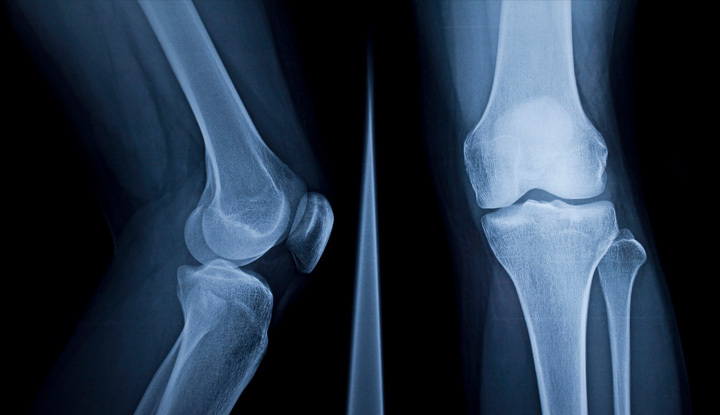
Image content: This image is available to view online.
View image online (https://my.clevelandclinic.org/-/scassets/images/org/health/articles/23501-knee-x-ray)
A knee X-ray is a test that produces a black-and-white image of the inside of your knee. The picture shows the soft tissues and bones in and around your knee joint. The bones include your kneecap (patella), part of your thigh bone (femur) and part of your shin bone (tibia). Part of your calf bone (fibula) can show up in knee X-rays too. Your healthcare provider may use knee X-rays to diagnose and treat health conditions involving your knee or knees.
Advertisement
Cleveland Clinic is a non-profit academic medical center. Advertising on our site helps support our mission. We do not endorse non-Cleveland Clinic products or services. Policy
X-rays use electromagnetic waves to create a picture of the inside of your body. X-rays are frequently the first type of imaging used when healthcare providers are trying to diagnose a condition. X-rays work by sending a beam of radiation through your body. Calcium in your bones absorbs more radiation, so your bones appear white on the X-ray. Soft tissues, such as muscle, fat and organs, absorb less radiation, so they appear different shades of gray.
Your healthcare provider may use a knee X-ray to diagnose possible health and medical conditions in your knee or knees. Knee X-rays can show signs of:
Knee X-rays are also used after a broken bone has been set to ensure your bone and/or joint has healed properly.
In addition, if you’ve had a knee replacement, your healthcare provider will want you to go in for routine follow-up X-rays to monitor your condition.
A radiologic technologist, also known as an X-ray technician, will perform your knee X-ray. Radiologic technologists must go through special training to learn about radiation, radiology and keeping people safe.
Advertisement
X-rays are a type of radiation. Small beams of radiation pass through your body and produce an image on special photographic film or a digital sensor.
Because your body parts have different degrees of thickness, they absorb different amounts of radiation. Bones, some tumors, calcifications and other solid matter show up white on X-rays. Less dense areas such as muscle and tissue allow radiation to pass through, so they look darker on X-rays.
You don’t have to do much to prepare for a knee X-ray. Make sure to wear comfortable clothing without any metal. You’ll be asked to remove any clothing, jewelry, belts or other objects containing metal. These may show up on the X-rays, interfering with getting a fully detailed image.
Tell your radiologic technologist if you’re pregnant or planning a pregnancy. Knee X-rays use a minimal amount of radiation, but there’s a chance your growing baby (fetus) could be exposed to it. Your healthcare provider will decide if you need the X-ray. If the X-ray is urgent, precautions will be taken to minimize radiation exposure to your baby.
Before your X-ray, your technologist will explain the X-ray process to you. If you have any questions about what’s going to happen, they’ll be happy to answer them for you.
A radiologic technologist will perform your knee X-ray in your healthcare provider’s office or a hospital radiology department. The procedure will take place in a room with a large X-ray machine hanging from the wall or ceiling. Once in the X-ray room, you may be given a lead apron to protect you from radiation exposure. The X-ray room is sometimes set at a cold temperature, but the process should only last about 10 minutes. An X-ray is like getting a picture of your knee taken — you can’t feel it, and it produces an image.
Your technologist will place an X-ray film holder or digital recording plate behind or under the X-ray machine. They’ll have you stand or sit in front of the X-ray machine or have you lie down on an X-ray table. You’ll need to keep very still during the procedure. Any movement may cause the X-ray images to show up blurry. The technologist may ask you to hold your breath while they’re taking the images.
Your technologist will go into a small room or behind a wall to operate the X-ray machine. They’ll return to reposition you for additional images. Sometimes, they may have you bend your knee or knees in certain ways as well.
A normal knee X-ray includes at least three different images. Your technologist will take one image from the front of your knee (anteroposterior view), one image from the side of your knee (lateral view) and one image of your kneecap with your knee bent (sunrise view). Sometimes, healthcare providers order X-rays of your other knee for comparison purposes or to look for signs of arthritis.
Advertisement
After your knee X-ray, your radiologic technologist will ensure that none of the images came out blurry. If any images need retakes, they’ll do them while you’re still there.
After that, a doctor called a radiologist will look at the X-ray images. Radiologists have special training in analyzing and interpreting X-rays. Once the radiologist has studied the results, they’ll send a report to your healthcare provider. Your healthcare provider will look over the results and discuss them with you. At that point, they’ll go over recommended treatment options.
Sometimes your healthcare provider will want additional images and you’ll have to return for follow-up X-rays. They’ll use these extra images to help make a correct diagnosis. You may also have to come back for a follow-up appointment to track your condition and watch for any changes that occur over time.
X-rays are a quick, easy way for your healthcare provider to diagnose a medical condition in your knee or knees. Knee X-rays contain very low amounts of radiation that go directly through your body. Also, X-rays typically don’t cause any side effects.
If you’re pregnant, your growing baby could be exposed to the slight radiation. Tell your radiologic technologist if you’re pregnant or thinking about getting pregnant. You may wear a lead apron to protect yourself and your baby from radiation exposure. Children have a slightly higher risk of issues with radiation exposure. Lower amounts may be used on children.
Advertisement
Extreme amounts of radiation exposure carry a small risk of cancer. However, the benefit of getting the correct diagnosis outweighs any risk of exposure. If you’re concerned about the amount of radiation you may be exposed to during an X-ray, talk to your technologist or healthcare provider.
Your healthcare provider may get immediate results if your knee X-ray was done due to an emergency. In other situations, your radiologist will have the results ready for your healthcare provider within two days. Your healthcare provider will discuss the findings with you and discuss treatment.
X-rays don’t clearly and accurately show your soft tissues such as ligaments, tendons and meniscus. To diagnose a tear in your ligaments, tendons or meniscus, your healthcare provider will order a computed tomography (CT) scan or a magnetic resonance imaging (MRI) scan. However, many orthopedic surgeons will request X-rays first to make sure they’re making the correct diagnosis.
If you have pain, swelling or tenderness in or around your knee or knees, your healthcare provider may order a knee X-ray. Knee X-rays can show signs of everything from fractures to infections to arthritis. Knee X-rays are quick and painless procedures that will help your healthcare provider diagnose you properly. And the sooner you’re diagnosed, the sooner you’ll be on your way to treatment.
Advertisement


Sign up for our Health Essentials emails for expert guidance on nutrition, fitness, sleep, skin care and more.
Learn more about the Health Library and our editorial process.
Cleveland Clinic’s health articles are based on evidence-backed information and review by medical professionals to ensure accuracy, reliability and up-to-date clinical standards.
Cleveland Clinic’s health articles are based on evidence-backed information and review by medical professionals to ensure accuracy, reliability and up-to-date clinical standards.
When you need a clear picture of what’s happening inside your body, the Cleveland Clinic imaging team is here for you.
