A brain (head) MRI scan is a painless test that produces very clear images of the structures inside of your head — mainly, your brain. Healthcare providers use brain MRIs to evaluate, diagnose and monitor several different medical conditions that affect your brain or other structures in your head.
Advertisement
Cleveland Clinic is a non-profit academic medical center. Advertising on our site helps support our mission. We do not endorse non-Cleveland Clinic products or services. Policy
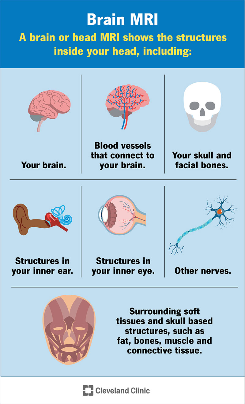
Image content: This image is available to view online.
View image online (https://my.clevelandclinic.org/-/scassets/images/org/health/articles/22966-brain-mri)
A brain MRI (magnetic resonance imaging) scan, also called a head MRI, is a painless procedure that produces very clear images of the structures inside of your head — mainly, your brain. MRI uses a large magnet, radio waves and a computer to produce these detailed images. It doesn’t use radiation.
Advertisement
Cleveland Clinic is a non-profit academic medical center. Advertising on our site helps support our mission. We do not endorse non-Cleveland Clinic products or services. Policy
Currently, MRI is the most sensitive imaging test of your head (particularly, your brain), as compared to other imaging techniques, such as CT (computed tomography) scans or X-rays.
Some brain MRI exams use an injection of contrast material. The contrast agent is often gadolinium, which is a rare earth metal. When this substance is present in your body, it alters the magnetic properties of nearby water molecules, which enhances the quality of the images. This improves the sensitivity and specificity of the diagnostic images.
Contrast material enhances the visibility of the following:
The contrast can also help diagnose multiple sclerosis, stroke, dementia and infection.
If your brain MRI requires a contrast material, your healthcare provider will insert an intravenous catheter (IV line) into a vein in your hand or arm. They’ll use this IV to inject the contrast material.
Contrast materials are safe intravenous (IV) drugs. Side effects, ranging from mild to severe, do occur, but severe reactions are very rare.
A head MRI and a brain MRI are the same procedure. They both provide images of the inside of your head. While healthcare providers most often use head and brain MRIs to assess your brain, these imaging procedures provide images of other structures in your head, too, such as facial bones, blood vessels and nerves.
Advertisement
A brain or head MRI shows the structures inside of your head, including:
More specifically, a brain or head MRI can show if there are any abnormalities in your brain or the surrounding tissues, including, but not limited to:
Neurologists and other healthcare providers order brain MRIs for several different reasons, including helping diagnose new neurological conditions based on certain symptoms or to monitor existing conditions.
Some of the conditions a brain MRI can help diagnose or monitor include:
Your healthcare provider may also order an MRI of your head if you have any combination of the following signs and symptoms:
Healthcare providers also use brain and head MRI scans before surgeries involving your head to better prepare for the surgery. They also use these scans to ensure that healing from the surgery is going well. Any significant injuries involving your head also prompt healthcare providers to order brain MRI scans to check for injuries, bleeding and swelling.
A radiologist or a radiology technologist will perform your brain (head) MRI. A radiologist is a medical doctor who performs and interprets imaging tests to diagnose conditions. A radiology technologist is a healthcare provider who’s specially trained and certified to perform an MRI scan.
Magnetic resonance imaging (MRI) works by passing an electric current through coiled wires to create a temporary magnetic field in your body — in this case, your head. A transmitter/receiver in the machine then sends and receives radio waves. The computer then uses these signals to make digital images of the structures inside of your head, including your brain.
Advertisement
Guidelines about eating and drinking before a brain MRI vary based on the reason for your MRI. Eat and take your medications as usual unless your healthcare provider tells you otherwise.
The magnetic resonance imaging (MRI) scanner uses strong magnets and radio wave signals that can cause heating or possible movement of some metal objects in your head and/or body. This could result in health and safety issues. It could also cause some implanted electronic medical devices to malfunction.
If you have metal-containing objects or implanted medical devices in your body, your healthcare provider needs to know about them before your brain MRI. Certain implanted objects may require additional scheduling arrangements and special instructions. Other items don’t require special instructions but may require an X-ray to check on the exact location of the object before your exam.
It’s important to tell your healthcare provider and MRI technologist if you have any of the following:
In addition, tell your healthcare provider if you:
Advertisement
Leave all jewelry and other accessories at home or remove them before your brain MRI. Metal and electronic items aren’t allowed in the exam room because they can interfere with the magnetic field of the MRI unit, cause burns or become harmful projectiles. These items include:
Most brain MRI exams are painless, but some people find it uncomfortable to remain still for 30 minutes or longer. Others may experience anxiety due to the closed-in space while in the MRI machine. The machine can also be noisy.
The general steps of a brain MRI scan and what to expect include:
Advertisement
In some cases, your MRI may require contrast. If this applies to you, your healthcare provider will give you an IV injection of contrast material before you undergo the MRI. The IV needle may cause some discomfort but this won’t last long. You may have some bruising afterward. Some people experience a temporary metallic taste in their mouth after the contrast injection.
If you have claustrophobia, your healthcare provider may recommend a sedative drug so you feel more relaxed during the exam, or even anesthesia.
In most cases, your whole body won’t go into the MRI machine tunnel if you’re only getting a head or brain MRI.
A brain MRI can take about 30 minutes to an hour to complete. It may take longer if you’re getting a brain MRI with contrast.
Your healthcare provider will be able to give you a more exact time range based on the specific reason for your scan.
After your MRI scan, a radiologist will analyze the images. The radiologist will send a signed report to your primary healthcare provider, who will share the results with you. The report is usually ready for your healthcare provider within one or two days.
You may need a follow-up exam. If so, your healthcare provider will explain why.
Brain magnetic resonance imaging (MRI) is a very useful and generally safe imaging test that healthcare providers use for a variety of reasons. If you need a brain MRI scan and are worried about the exam or have questions about it, don’t hesitate to ask your healthcare provider. They’re available to answer your questions and support you.


Sign up for our Health Essentials emails for expert guidance on nutrition, fitness, sleep, skin care and more.
Learn more about the Health Library and our editorial process.
Cleveland Clinic’s health articles are based on evidence-backed information and review by medical professionals to ensure accuracy, reliability and up-to-date clinical standards.
Cleveland Clinic’s health articles are based on evidence-backed information and review by medical professionals to ensure accuracy, reliability and up-to-date clinical standards.
If you have a neurological condition, you want expert advice. At Cleveland Clinic, we’ll work to create a treatment plan that’s right for you.
