A HIDA scan is an imaging procedure that shows how well your liver, bile ducts and gallbladder are functioning. It can help diagnose certain conditions, such as cholecystitis, biliary leak and biliary atresia.
Advertisement
Cleveland Clinic is a non-profit academic medical center. Advertising on our site helps support our mission. We do not endorse non-Cleveland Clinic products or services. Policy
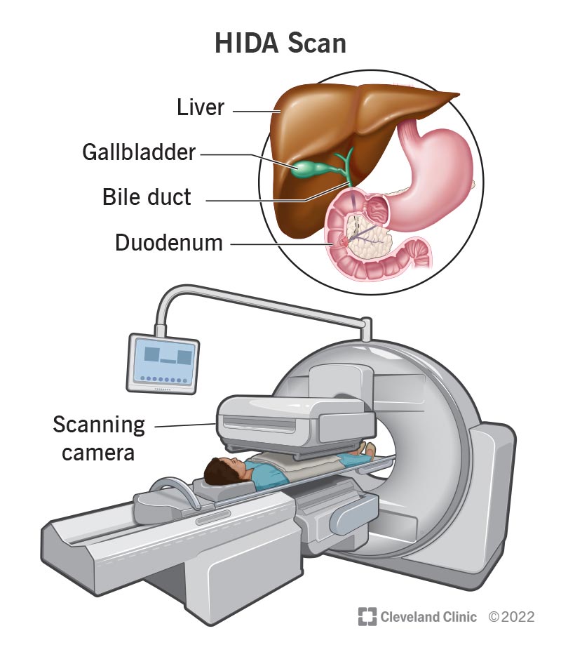
Image content: This image is available to view online.
View image online (https://my.clevelandclinic.org/-/scassets/images/org/health/articles/17099-hilda-scan)
A HIDA scan (hepatobiliary iminodiacetic acid scan) is an imaging procedure that uses an injected chemical called a radioactive tracer (radiotracer) and a scanning camera to evaluate your gallbladder. The scan is performed in the department of nuclear medicine in radiology.
Advertisement
Cleveland Clinic is a non-profit academic medical center. Advertising on our site helps support our mission. We do not endorse non-Cleveland Clinic products or services. Policy
More specifically, the HIDA scan tracks the flow of bile from your liver to your small intestine. This is known as the biliary system.
Your liver makes bile (digestive fluid) that helps your body break down the fat in the food you eat. Certain ducts (biliary ducts) carry bile from your liver to your gallbladder for storage. Your gallbladder is a small sac under your liver on your right side at the level of your lower ribs. When you eat food, your gallbladder contracts (squeezes) and releases stored bile through ducts into the first part of your small intestine (the duodenum) to help break down the fats.
If any part of this process isn’t working properly, it can cause certain symptoms and conditions, which a HIDA scan can help diagnose.
Other names for a HIDA scan include cholescintigraphy and hepatobiliary scintigraphy.
Healthcare providers use a HIDA scan to help diagnose and evaluate the following conditions:
Advertisement
Healthcare providers perform HIDA scans to evaluate conditions that affect liver cells, the ducts of the biliary system and your gallbladder.
Your provider may recommend a HIDA scan if you have any of the following symptoms:
You may also need a HIDA scan if you’ve had a biliary stent placed. A biliary stent (bile duct stent) is a thin, hollow tube that’s placed in the bile ducts. The stent holds the duct open if it’s been blocked or partly blocked. Your provider may request a HIDA scan to make sure the stent is working properly.
If you’ve had a liver transplant, you may need multiple HIDA scans to evaluate the function of your liver after the transplant surgery.
Three medical professionals are involved in performing a HIDA scan to calibrate the scanning equipment, provide radiopharmaceuticals (radiotracers) and interpret the results of the scan. These providers include:
A HIDA scan (hepatobiliary iminodiacetic acid scan) uses small amounts of radioactive substances called radiopharmaceuticals or radiotracers that a healthcare provider typically injects into your bloodstream.
The radiotracer then travels through your liver and into your gallbladder and your small intestines. The radiotracer gives off energy in the form of gamma rays. Special cameras detect this energy and, with the help of a computer, create detailed pictures that show how your organs and tissues function.
Your healthcare team will give you specific instructions to prepare for a HIDA scan. Be sure to follow them. Here are some general guidelines to prepare for a HIDA scan:
Advertisement
HIDA scans don’t typically require anesthesia to put you to sleep (or to prevent pain). In fact, for some scans, you may need to move into different positions.
If you may have issues remaining still during the scan or if your newborn or child is getting the scan, you or your child may be given medicine (a sedative) that makes you relaxed and sleepy — but still awake —during the scan.
A HIDA scan usually takes one to four hours. In some cases, you may need to return for additional imaging up to 24 hours after the first scan.
A HIDA scan procedure can have slightly different steps depending on which part of your biliary system your healthcare provider is evaluating.
In general, you can expect the following during a HIDA scan:
Advertisement
The HIDA scan itself is painless. If you receive the radiotracer through an IV, you may feel a brief sting or pinch as your provider places the IV in your arm.
However, you may be in pain while undergoing a HIDA scan because of the condition your provider is trying to diagnose. For example, cholecystitis and sphincter of Oddi dysfunction often cause severe pain. And you may not be able to be on pain medication for the scan because some medications alter the function of your biliary system and would interfere with the accuracy of the test.
Opiates (like morphine and codeine), for example, need to be withheld for at least six hours before a HIDA scan.
Depending on the reason for your HIDA scan, you may be able to go home or you’ll return to your hospital room.
Be sure to drink lots of fluids for the next 24 hours after your scan to help flush the radiotracer out of your body. Most of the radiotracer will leave your body through your urine or stool within a day.
Be sure to flush the toilet right after you use it, and wash your hands thoroughly with soap and water. The amount of radiation in the tracer is very small, so it isn't a risk for people to be around you after the scan.
If you’re breastfeeding, you’ll need to discard the milk you pump for 24 hours after the scan. This is because your breastmilk can have radiation in it from the radiotracer, which can harm your baby. You may wish to pump additional breast milk prior to the scan and safely store it or make alternate plans for your baby to receive nutrition for the one day after the scan.
Advertisement
A HIDA scan has very few risks, including:
It’s important to tell your healthcare provider if you’re pregnant, think you might be pregnant or are breastfeeding. In most cases, providers don’t perform nuclear medicine tests, such as the HIDA scan, on someone who's pregnant due to potential harm to the developing fetus.
Nuclear medicine imaging, which includes a HIDA scan, provides unique information that providers can’t often get using other imaging procedures, such as ultrasound. Because of this, the benefits of a HIDA scan far outweigh the risks if you're not pregnant.
A radiologist will interpret the images of the HIDA scan, write a report and share the results with your healthcare provider. Your provider will then share the results with you. This process usually takes less than 24 hours.
The results report for your HIDA scan may have different information depending on why you needed the scan. In general, the report will detail how the radiotracer flowed through your biliary system.
Healthcare providers use the results of a HIDA scan and other testing, such as blood tests, to make a final diagnosis.
The possible report findings for a HIDA scan may include:
It’s important to remember that your provider will explain the results to you, whatever they may be. Feel free to ask them questions.
A HIDA scan (hepatobiliary iminodiacetic acid scan) is an important test for diagnosing certain issues in your liver, bile ducts and gallbladder. While it can be stressful to have to undergo a test, know that a HIDA scan is essentially painless and that your healthcare provider is available to answer any questions or concerns you may have.


Sign up for our Health Essentials emails for expert guidance on nutrition, fitness, sleep, skin care and more.
Learn more about the Health Library and our editorial process.
Cleveland Clinic’s health articles are based on evidence-backed information and review by medical professionals to ensure accuracy, reliability and up-to-date clinical standards.
Cleveland Clinic’s health articles are based on evidence-backed information and review by medical professionals to ensure accuracy, reliability and up-to-date clinical standards.
When you need a clear picture of what’s happening inside your body, the Cleveland Clinic imaging team is here for you.
