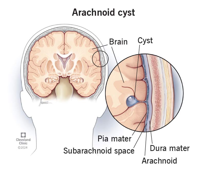
An arachnoid cyst is a noncancerous fluid-filled sac that grows on your brain or spinal cord. Most of the time, it’s something you were born with. It’s rare to cause symptoms or need treatment. But if you have a large or changing cyst, your provider might recommend surgically removing or draining it.
Advertisement
Cleveland Clinic is a non-profit academic medical center. Advertising on our site helps support our mission. We do not endorse non-Cleveland Clinic products or services. Policy

Image content: This image is available to view online.
View image online (https://my.clevelandclinic.org/-/scassets/images/org/health/articles/arachnoid-cyst)
Arachnoid cysts are fluid-filled sacs that form on your brain or spinal cord. They’re filled with cerebrospinal fluid. This surrounds and protects your central nervous system (CNS).
Advertisement
Cleveland Clinic is a non-profit academic medical center. Advertising on our site helps support our mission. We do not endorse non-Cleveland Clinic products or services. Policy
These cysts form when the arachnoid membrane tears. This membrane is a thin, spiderweb-like layer that cushions your brain and spinal cord.
Most arachnoid cysts never cause symptoms. But if they grow large (which is rare), they can press on and damage parts of your CNS.
Your brain and spinal cord are covered by a thin layer of tissue called the arachnoid membrane. Arachnoid cysts form under this protective layer.
Intracranial arachnoid cysts are ones that grow on your brain. They can form in different areas, including:
Spinal arachnoid cysts grow on your spinal cord. They’re most common in your thoracic spine. This runs from your neck to the bottom of your ribs. They’re much less common than intracranial arachnoid cysts.
There are two types based on when they form:
Most arachnoid cysts don’t cause symptoms (asymptomatic). They usually only cause problems if they grow large enough to press on your brain or spinal cord. Symptoms depend on the location, size and whether they press on nerves.
Possible symptoms include:
Advertisement
An arachnoid cyst forms when the arachnoid membrane splits or tears. Fluid around the area collects in a sac.
Healthcare providers don’t know exactly why primary arachnoid cysts are present at birth. They may form if the membrane tears while the fetus is growing.
Secondary cysts can develop after:
Because the cause is often unknown, you usually can’t prevent these cysts.
Anyone can develop a cyst. But they’re most common in babies and young children. Males are more likely to get them.
You may also be more at risk if you have certain health conditions, like arachnoiditis or Marfan syndrome.
Sometimes, arachnoid cysts run in families, so there could be a genetic link.
If you’re concerned about passing the risk to your children, talk to a healthcare provider about genetic counseling. They may suggest genetic testing.
Complications are rare. Children born with a cyst may need more time to meet developmental milestones for their age, like taking their first steps. Cysts can also affect hormone production that targets their growth, sexual development and metabolism.
You might notice the following if an arachnoid cyst grows too large for the area it’s in:
Cysts may block the flow of cerebrospinal fluid around your brain. This is called hydrocephalus. It’s dangerous because it can increase pressure in your skull.
A cyst can also break open (rupture). This can release extra CSF into your brain or spinal cord. This is a medical emergency. It can cause sudden pressure, bleeding or fluid buildup.
A healthcare provider diagnoses an arachnoid cyst with a physical exam and imaging tests. If you or your child has symptoms, they’ll ask about your health history.
You may need an MRI or CT scan to take pictures of the cyst.
Most providers find arachnoid cysts by accident. This happens if your provider runs tests for another reason and finds the cyst during that time. Because most cysts don’t cause symptoms, you may have one for a long time without knowing it.
You probably won’t need any treatment if you have a cyst. Most of them never cause symptoms or get big enough to cause damage. Your provider may recommend regular MRIs or CT scans to see if the cyst grows or changes.
Advertisement
But if you have symptoms, your provider may suggest surgery.
If the cyst is large, growing or putting pressure on your brain, spinal cord or nerves, your provider may recommend surgery. There are different options:
The best treatment for you depends on the cyst’s location and size. Your provider and surgeon will explain what to expect and how long recovery will take.
Advertisement
See a provider right away if you or your child has symptoms of an arachnoid cyst. Some cysts need quick treatment. They can share symptoms with serious conditions like brain tumors, so make sure a healthcare provider checks your symptoms to rule out anything serious.
Get medical help immediately if you notice sudden changes in how you move, think, see or feel.
Most arachnoid cysts don’t cause problems. Rarely, a hard hit could make it break open. This requires medical care. Wear safety gear, like a helmet, when needed, to prevent any accidents and injuries.
In some cases, you may need treatment. Removing the whole cyst can cure it. If your surgeon can’t safely remove it all, it can fill up with fluid again. Sometimes, an arachnoid cyst can grow back after a complete removal. But this isn’t common.
If your provider found the cyst by accident while checking for something else, you usually don’t need to worry. They may watch it over time to see if it gets bigger or moves.
If you just found out there’s a growth in your brain or spinal cord, your mind is probably racing. But you may have sigh in relief if the growth is an arachnoid cyst. That’s because most arachnoid cysts are harmless. Many of them never need treatment.
Advertisement
Keep in touch with your provider. They’ll tell you how often you’ll need follow-up tests and appointments to keep an eye on the cyst.
While arachnoid cysts are rarely serious, you may still have lots of questions. Don’t be afraid to ask your provider about anything that crosses your mind. There’s no such thing as a silly question when it comes to your brain or spine health.
Learn more about the Health Library and our editorial process.
Cleveland Clinic's health articles are based on evidence-backed information and review by medical professionals to ensure accuracy, reliability, and up-to-date clinical standards.
Cleveland Clinic's health articles are based on evidence-backed information and review by medical professionals to ensure accuracy, reliability, and up-to-date clinical standards.
Cleveland Clinic’s primary care providers offer lifelong medical care. From sinus infections and high blood pressure to preventive screening, we’re here for you.
