If you or someone you love needs vision correction but can’t communicate or participate in an eye exam, retinoscopy can help. This technique relies on optics (the science of how light behaves) and eye anatomy. Eye specialists can use retinoscopy to help anyone needing vision correction, not just those with special needs and circumstances.
Advertisement
Cleveland Clinic is a non-profit academic medical center. Advertising on our site helps support our mission. We do not endorse non-Cleveland Clinic products or services. Policy
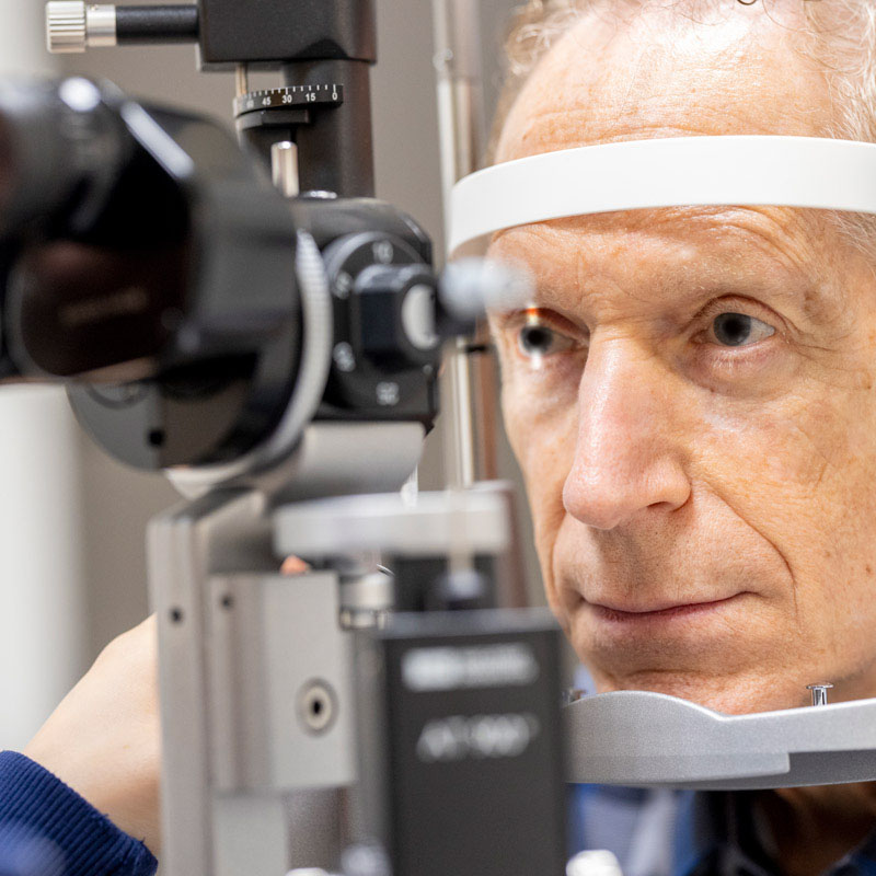
Image content: This image is available to view online.
View image online (https://my.clevelandclinic.org/-/scassets/images/org/health/articles/retinoscopy)
Retinoscopy is an eye test that lets an eye care specialist find and measure refractive errors using special handheld tools and light that reflects off the retinas inside your eyes. While advances in technology mean eye specialists have other ways to measure refractive errors, this technique remains extremely common and it’s frequently a part of routine eye exams.
Advertisement
Cleveland Clinic is a non-profit academic medical center. Advertising on our site helps support our mission. We do not endorse non-Cleveland Clinic products or services. Policy
Retinoscopy is especially useful for eye tests on people who can’t communicate with their eye specialists. Have you ever wondered how infants or people with language difficulties and barriers can get prescription vision correction? The answer is retinoscopy.
In addition to very young children and people who can’t speak the same language as their eye specialists, retinoscopy is also vital for people with intellectual difficulties or age-related brain diseases. There’s also a subtype of retinoscopy that lets your specialist diagnose trouble focusing your eyes at different distances.
Retinoscopy relies on how light travels through the corneas and lenses of your eyes and reflects off your retinas. If the light entering your eyes is bright and direct enough, the reflection causes a glow in your pupils that’s visible to others. Experts call that “fundus reflex” or “ “red reflex.”
When you have an eye exam, a key part of that exam is testing your visual acuity to see if you have normal 20/20 vision. When you have 20/20 vision, light rays bouncing off your retinas should line up so they’re parallel when they leave your eyes. But if you have a refractive error, the error bends those rays differently, causing some of them to misalign. Retinoscopy lets an eye care specialist find which vision corrections make the light rays align correctly. Those corrections become your prescription.
Advertisement
Retinoscopy can also help with diagnosing refractive errors when you have eye conditions like:
In most cases, you won’t need to prepare for retinoscopy (either on its own or if it’s part of a broader eye exam). There are a few exceptions, but these are usually very specific. Your eye specialist will tell you if this is the case before your exam. They’ll also let you know how long before your exam you’ll need to stop wearing your contacts.
To do standard retinoscopy, an eye care specialist first has to use eyedrops that contain cycloplegic medications. Those medications keep your eyes from trying to automatically focus during this test, which makes it harder for your eye specialist to determine your prescription.
If your provider is going to do dynamic retinoscopy, they won’t use cycloplegic medications before doing it. That test relies on your eyes’ natural focusing reflex.
During retinoscopy, your eye specialist will have you seated in an exam room with the lights dimmed. You’ll sit in front of them, and you’ll be facing each other. One of the tools they’ll use is a retinoscope. This is a handheld tool that looks like someone combined a heavy-duty flashlight and a magnifying glass. The magnifying window and light on the top of the retinoscope are set up so your eye specialist can shine the light into your eye and look closely at your eye at the same time.
The other key part of retinoscopy is a way to test different lens strengths and how they affect the reflected light in your pupils. There are two main ways they can do this:
When your eye specialist shines their light through each lens into your eye, they’re watching your pupils for specific changes in the reflected glow. Those changes include:
Advertisement
When your eye specialist shines their light through a lens and into your eye, they’ll move the light up and down, left and right, and diagonally several times. They’ll also change out the lenses they use while they do this. Your provider will probably repeat these actions several times and will stop at different points to adjust the light on the retinoscope or take notes.
One specific approach to retinoscopy tests your eyes’ ability to focus up close. When your eyes focus up close, that’s called “accommodation.” An eye specialist can test this ability using a technique called dynamic retinoscopy.
If your specialist is using dynamic retinoscopy to test your eyes’ focusing ability, the procedure is similar but may not involve magnifying lenses. Instead, your specialist will have you look at objects at different distances. Some of those objects will be between you and your specialist, while others will be behind your specialist (but still visible just to their side or over their shoulder).
While you look at these objects, your provider watches the glow in your pupils from their retinoscope’s light. The way the glow moves lets your provider judge if your eyes’ accommodation ability works as it should.
Advertisement
If your provider used cycloplegic medication to dilate your pupils or block your eyes’ accommodation reflexes, your eyes will stay dilated for a while after your exam. The time it takes for these medications to wear off varies. Your eye specialist can tell you about how long you should expect that to take.
Your eye specialist may also give you temporary sunshades or a shading cover to wear over your glasses. Cycloplegic medications also paralyze your pupils, which means they can’t narrow in bright conditions to reduce how much light enters your eyes.
Retinoscopy has almost no risks or side effects. There are some side effects that cycloplegic mediations can cause, but these are usually minor. And some people who are sensitive to bright light may find parts of retinoscopy less pleasant, so if you have light sensitivity (photophobia), be sure to tell your eye specialist at the start of your exam. Your eye specialist can also answer any questions or concerns about cycloplegic medication side effects.
After your eye care specialist finishes the retinoscopy test, they’ll calculate your vision prescription (or the prescription of your loved one who was undergoing this test). The prescription works the same way as a prescription from a subjective refraction exam. Your specialist can tell you about your prescription, including what it means for you.
Advertisement
If you or your loved one doesn’t have normal 20/20 vision, your (or their) eye specialist will talk to you about prescription vision correction. The most common forms of correction are eyeglasses or contact lenses, but vision correction surgeries like LASIK are also available for some people.
A good vision correction prescription is like a tailored piece of clothing that fits you perfectly, and an eye care specialist using retinoscopy is like a master tailor sewing every stitch by hand so you get that perfect fit. Though this technique is over 150 years old, it remains common today and sees frequent use in routine eye exams. Whether it’s for you or for someone you love, retinoscopy is one way you can feel reassured that your eye specialist can give you the best possible prescription for your needs.


Sign up for our Health Essentials emails for expert guidance on nutrition, fitness, sleep, skin care and more.
Learn more about the Health Library and our editorial process.
Cleveland Clinic’s health articles are based on evidence-backed information and review by medical professionals to ensure accuracy, reliability and up-to-date clinical standards.
Cleveland Clinic’s health articles are based on evidence-backed information and review by medical professionals to ensure accuracy, reliability and up-to-date clinical standards.
Cleveland Clinic’s ophthalmologists and optometrists have the highest training available. We provide exams, vision correction and care for many eye conditions.
