Looking inside a person’s eyes is a tried-and-true method for gauging a person’s overall health. And fundoscopy is a test that medical providers of all backgrounds and specialties can use to take in that view. This simple, painless test is a key tool in many types of medical examinations, routine and otherwise.
Advertisement
Cleveland Clinic is a non-profit academic medical center. Advertising on our site helps support our mission. We do not endorse non-Cleveland Clinic products or services. Policy
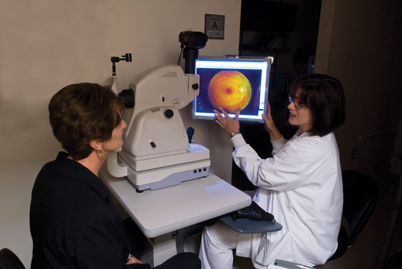
Image content: This image is available to view online.
View image online (https://my.clevelandclinic.org/-/scassets/images/org/health/articles/fundoscopy)
Fundoscopy is a simple exam where a healthcare provider uses magnifying tools with an attached light to look into your eyes. It’s a common part of many routine medical exams. And it’s also a common part of diagnosing illnesses, injuries and many other conditions.
Advertisement
Cleveland Clinic is a non-profit academic medical center. Advertising on our site helps support our mission. We do not endorse non-Cleveland Clinic products or services. Policy
The term gets its name from the word “fundus,” the medical term for the inner back wall of your eye. Fundoscopy is also commonly known as ophthalmoscopy (pronounced “off-thal-MOSS-co-pea”) because an ophthalmoscope is one of the tools most often used for it.
There are two types of fundoscopy that a provider can perform, with a specific device for each type:
There’s also a variant of indirect ophthalmoscopy where a provider uses a handheld ophthalmoscope in one hand that they look through with one eye while using their other hand to use a condensing lens. This variant is called monocular indirect ophthalmoscopy. It doesn’t have the advantage of 3D vision and depth perception, though.
Advertisement
Fundoscopy (pronounced "fun-DOSS-ko-pea") is a routine part of medical visits of many kinds. It’s especially common with seeing primary care providers or children’s visits to pediatricians. It can also be part of the following:
Providers can also take this test a step further and do what’s called “extended ophthalmoscopy.” Doing that involves taking a closer view and sketching or drawing what the back of your eye looks like. It’s more common after retinal surgeries or with specific types of retinal conditions.
The pupil of your eye is actually just a window-like opening. The iris, a ring of muscular tissue around the pupil that gives you your eye color, controls how wide the pupil is. Light entering your eye through your pupil is one of the earliest steps in how your vision works.
Healthcare providers can take advantage of how your eye works by using an ophthalmoscope to look into your eyes. The light on the ophthalmoscope illuminates the inside of your eye, and magnifying lenses give your provider a more detailed view.
When using an ophthalmoscope, your provider is looking for specific things that should be visible at the back of your eye. The things they’re trying to see include your:
Fundoscopy usually doesn’t take any preparation on your part (your provider or eye specialist can tell you if there are any exceptions to that).
In some cases, fundoscopy happens after a provider or eye specialist gives you eye drops to dilate your pupils. This is especially common when fundoscopy is part of an eye exam.
During fundoscopy, a healthcare provider or eye specialist will have you seated in front of them or lying down. This often happens in rooms with dimmed lights to make it easier to see into your eyes. They’ll have you look straight ahead or occasionally have you turn your eyes to look in various directions. If they’re doing indirect ophthalmoscopy, they’ll also hold a handheld lens just in front of your eye. They’ll then align the handheld lens so they can see through it and the viewing window in the ophthalmoscope.
Eye dilation is often part of this test, especially if it’s happening during an eye exam. Dilation lets your provider get a wider view inside your eyes, which can make it easier to see smaller or otherwise hidden issues. They can also tell you about what they’re looking for and what — if anything — they see that might be a cause for concern.
Advertisement
If they see anything that might be a cause for concern, they may also recommend an additional test like fundus photography or a slit lamp exam. It varies, so don’t hesitate to ask your provider or eye specialist more about any tests they recommend and what their concerns might be.
Your provider or eye specialist can tell you what they saw inside your eyes. That includes explaining any potential causes for concern. They can also offer guidance on what you might need to do or understand moving forward.
Some providers may choose to talk you through the process as they’re doing it, giving you a play-by-play description. Other providers may choose to focus on the exam and tell you what they saw in more detail afterward. For the most part, how they tell you is a personal preference. But if you’re anxious or worried, you can tell your provider that before they start doing the fundoscopy exam. They can take your concerns and feelings into account to help reassure you and ease those concerns.
The eyes are the window to the soul, as the saying goes. But they’re also a window to your health. Medical professionals from many specialties and backgrounds use fundoscopy to look into your eyes and gauge the health of your eyes, brain and body. Fundoscopy (also known as ophthalmoscopy) is one of the most important ways for providers to look into your eyes for important clues or changes that could indicate an issue that needs care.
Advertisement
Better still, this test is fast, painless and can even be done in ways that involve minimal equipment costs — helping make it available worldwide, even in areas with limited access to medical care and technology.
Advertisement


Sign up for our Health Essentials emails for expert guidance on nutrition, fitness, sleep, skin care and more.
Learn more about the Health Library and our editorial process.
Cleveland Clinic’s health articles are based on evidence-backed information and review by medical professionals to ensure accuracy, reliability and up-to-date clinical standards.
Cleveland Clinic’s health articles are based on evidence-backed information and review by medical professionals to ensure accuracy, reliability and up-to-date clinical standards.
Cleveland Clinic’s ophthalmologists and optometrists have the highest training available. We provide exams, vision correction and care for many eye conditions.
