A breast ultrasound is an imaging test your provider uses to get pictures of the inside of your breast. You might need a breast ultrasound if your provider wants another look at an area that was abnormal or unclear on a mammogram. Your provider might also recommend getting a breast ultrasound in addition to a mammogram for breast cancer screenings.
Advertisement
Cleveland Clinic is a non-profit academic medical center. Advertising on our site helps support our mission. We do not endorse non-Cleveland Clinic products or services. Policy
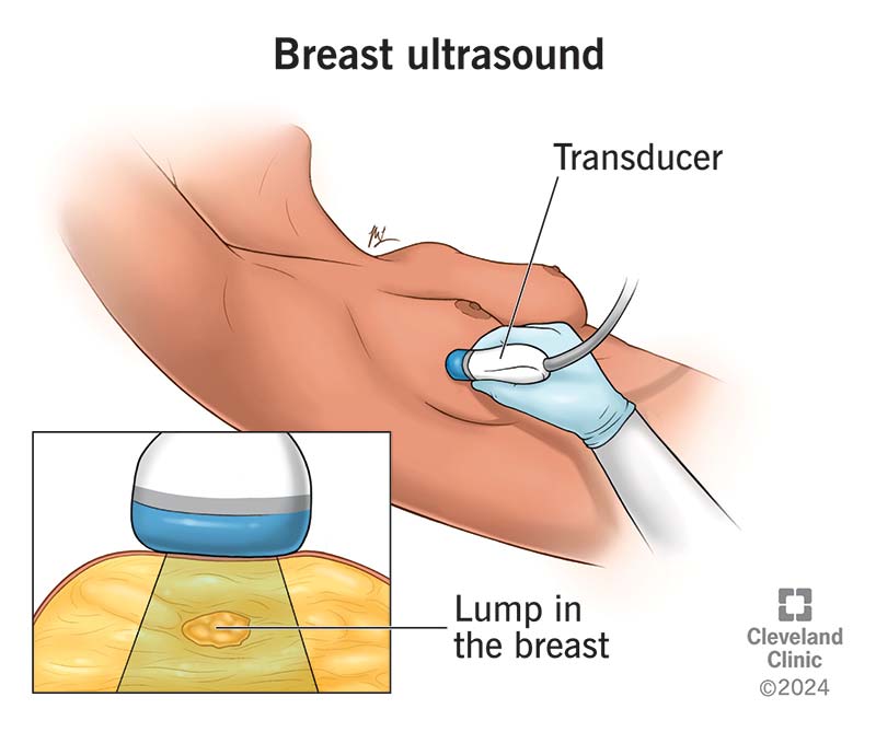
Image content: This image is available to view online.
View image online (https://my.clevelandclinic.org/-/scassets/images/org/health/articles/21496-breast-ultrasound)
A breast ultrasound is an imaging test that healthcare providers use to take pictures of the inside of your breast. It can allow your provider to focus on a small area of your breast that they need to see more closely. Providers might have you get one after a mammogram to get a detailed look at changes they see, or in addition to a mammogram to screen for breast cancer.
Advertisement
Cleveland Clinic is a non-profit academic medical center. Advertising on our site helps support our mission. We do not endorse non-Cleveland Clinic products or services. Policy
A breast ultrasound can show whether a breast lump is a fluid-filled breast cyst (usually not cancerous) or a solid mass (which could be cancer and may need further testing).
Your healthcare provider might order a breast ultrasound if:
Providers can also use a breast ultrasound during a biopsy to make sure they take a sample of tissue from the right spot. A pathologist can then look at the tissue under a microscope to diagnose or rule out breast cancer.
To get images of the inside of your breast, an ultrasound technician moves a handheld device (transducer) over your breast and surrounding tissues. The transducer uses high-frequency sound waves to take pictures of the tissues and structures inside. The technician uses a computer monitor to look at the images the transducer collects.
Advertisement
On the day of your ultrasound, don’t wear deodorant or use lotions or powders on or around your breasts. These products can make it hard to get clear images. You also might want to wear comfortable clothing so it’s easier to change.
During a breast ultrasound:
A breast ultrasound takes about half an hour.
When the ultrasound is complete, you or the technician will wipe any remaining gel off your skin. A radiologist will look at and interpret the images. Your provider will tell you if you need any follow-up tests or procedures. Sometimes, the provider will recommend a biopsy based on the images. They’ll schedule the biopsy before you leave.
Breast ultrasounds create images with sound waves, so there’s no exposure to radiation. There are no known risks of ultrasound technology. But it’s limited — providers usually use it to see one specific area of your breast. That’s why mammography is still the best tool for looking at your entire breast. If you’re at an increased risk of breast cancer, your provider may recommend a breast ultrasound in combination with a mammogram for screening.
You should know the results of your breast ultrasound by the end of your appointment. Your provider will let you know if what they see on the ultrasound looks noncancerous (benign), like a cyst, or is potentially cancerous (malignant). They’ll schedule or perform follow-up procedures right away.
Call your healthcare provider if you:
If you need a breast ultrasound, your mind might go to the worst-case scenario. You may feel vulnerable and anxious about the process. But know that a breast ultrasound is a safe, noninvasive way for a provider to get a better look at a specific area of your breast. Your technician will help make you as comfortable as possible.
Advertisement
Many lumps aren’t cancerous. But no matter what the results are, your healthcare team will help you through the next steps.
Advertisement


Sign up for our Health Essentials emails for expert guidance on nutrition, fitness, sleep, skin care and more.
Learn more about the Health Library and our editorial process.
Cleveland Clinic’s health articles are based on evidence-backed information and review by medical professionals to ensure accuracy, reliability and up-to-date clinical standards.
Cleveland Clinic’s health articles are based on evidence-backed information and review by medical professionals to ensure accuracy, reliability and up-to-date clinical standards.
A mammogram is a way to keep track of changes in your breasts. Cleveland Clinic’s experts can help you get a mammogram and connect you to the care you need.
