Your cerebral cortex, also called gray matter, is your brain’s outermost layer of nerve cell tissue. It has a wrinkled appearance from its many folds and grooves. Your cerebral cortex plays a key role in memory, thinking, learning, reasoning, problem-solving, emotions, consciousness and functions related to your senses.
Advertisement
Cleveland Clinic is a non-profit academic medical center. Advertising on our site helps support our mission. We do not endorse non-Cleveland Clinic products or services. Policy
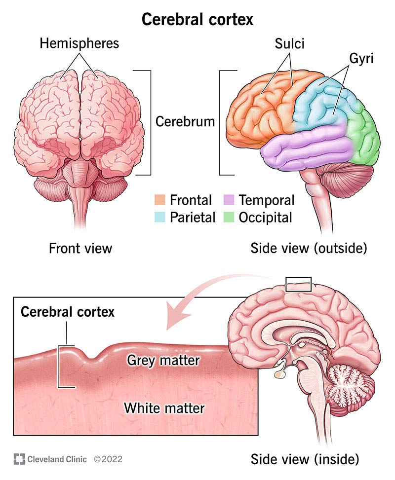
Image content: This image is available to view online.
View image online (https://my.clevelandclinic.org/-/scassets/images/org/health/articles/23073-cerebral-cortex)
Your cerebral cortex is the outermost layer of your brain. Its surface has many folds, giving it a wrinkled appearance. The folds consist of many deep grooves called sulci and raised areas called gyri. These folds add to the surface area of your cerebral cortex, allowing large amounts of information to be processed by more nerve cells. Your cerebral cortex makes up about half of your brain’s total mass.
Advertisement
Cleveland Clinic is a non-profit academic medical center. Advertising on our site helps support our mission. We do not endorse non-Cleveland Clinic products or services. Policy
Your cerebral cortex consists of six layers of nerve cells that contain between 14 billion and 16 billion nerve cells. It’s two millimeters (mm) to four mm (0.08 inches to 0.16 inches) thick.
Your cortex is divided into four lobes: frontal, parietal, temporal and occipital. Each of these lobes is responsible for processing different types of information. Collectively, your cerebral cortex is responsible for the higher-level processes of the human brain, including language, memory, reasoning, thought, learning, decision-making, emotion, intelligence and personality.
The gray matter found in the outer layer of your brain consists of nerve cell bodies, including the end portion of nerves called dendrites. Dendrites are the part of a nerve cell that receives the chemical message from another cell. Your cerebral cortex is gray because that section of the nerve lacks the fatty covering material called myelin.
White matter in your brain is made up of bundles of axons, the long center section of a nerve cell that’s wrapped in myelin. The myelin gives the tissue its whitish color.
Your cerebral cortex is the outer layer that lies on top of your cerebrum. Your cerebrum is the largest area of your brain. Your cerebrum divides your brain into two halves called hemispheres. The hemispheres are attached by a bundle of nerve fibers called the corpus callosum. The corpus callosum allows your two hemispheres to communicate with each other.
Advertisement
Most of your cerebral cortex is considered to be the neocortex. “Neo” means new. Your neocortex is so named because its appearance is thought to be relatively new in vertebrate evolution. In humans, 90% of the cerebral cortex is the neocortex.
Your cerebral cortex is involved in many high-level functions, such as reasoning, emotion, thought, memory, language and consciousness. Each lobe of your brain is associated with different functions.
Your frontal lobe is at the front of your brain behind your forehead. Functions of your frontal lobe include:
Special areas of note within this lobe are the motor cortex, the prefrontal cortex and Broca’s area. Your motor cortex is responsible for body movement. Your prefrontal cortex is in charge of “executive functions,” such as thinking and problem-solving. It also supervises and directs other areas of your brain. Broca’s area is a part of your frontal lobe that’s involved with speech production.
Your occipital lobe is at the back of your brain. Functions of your occipital lobe include:
Your parietal lobe is located between your frontal and occipital lobes and above your temporal lobe. Functions of your parietal lobe include:
Special areas of note within this lobe are the somatosensory cortex. It receives sensory information (“feeling” information) from all over your body. Here’s an example of how brain lobes work together:
The motor cortex in your brain’s frontal lobe sends the message that directs the muscles in your arm and hand to reach out toward a cup of soup on your kitchen table. The somatosensory cortex of your parietal lobe assesses the information delivered through your touch of the cup, including judgment of its temperature. Spatial processing in your parietal lobe allows you to grasp the cup, flawlessly navigating hand-to-cup distance relative to the table and other surrounding objects.
Your temporal lobe is located between your frontal and occipital lobes and below your parietal lobe. Functions of your temporal lobe include:
Advertisement
A special area of note within this lobe is Wernicke’s area. This area was more recently discovered to be involved in language comprehensive based on speech tones and sounds, linking them to previously learned sounds.
Some researchers look at the brain in another way and classify the areas of the cerebral cortex by their three main types of functions: sensory, motor and association areas.
Sensory areas: These areas of your cerebral cortex receive sensory information from your senses and your environment. Functions include:
Advertisement
Motor areas: These areas of your cerebral cortex are involved in voluntary muscle movement. These functions are processed mainly by your frontal lobe. Functions include:
Association areas: These areas are spread throughout all four lobes and connect and add complexity to functions. Functions include:
Damage to any area of your cerebral cortex typically results from tumors, trauma, autoimmune diseases or a cerebrovascular accident (brain bleed or stroke).
Symptoms depend on the area of the cerebral cortex that’s damaged.
Symptoms of damage or injury to your frontal lobe include:
Advertisement
An additional cause of damage to the frontal lobe is dementia.
Symptoms of damage to your parietal lobe include:
Symptoms of damage to your temporal lobe include:
Additional causes of damage to the temporal lobe include epileptic seizures, developmental dyslexia and Alzheimer’s disease.
Symptoms of damage to your occipital lobe include:
Your cerebral cortex is the outer covering of the surface of your brain. It consists of between 14 billion and 16 billion nerve cells. Your cortex is involved in higher processes in the human brain, including memory, thinking, learning, reasoning, problem-solving, emotions, consciousness and functions related to your senses.


Sign up for our Health Essentials emails for expert guidance on nutrition, fitness, sleep, skin care and more.
Learn more about the Health Library and our editorial process.
Cleveland Clinic’s health articles are based on evidence-backed information and review by medical professionals to ensure accuracy, reliability and up-to-date clinical standards.
Cleveland Clinic’s health articles are based on evidence-backed information and review by medical professionals to ensure accuracy, reliability and up-to-date clinical standards.
If you have a neurological condition, you want expert advice. At Cleveland Clinic, we’ll work to create a treatment plan that’s right for you.
