Equipment
Our computing facilities are housed in our laboratory space and distributed across a 100 megabit-per-second fast ethernet LAN. It consists of a 6-node Linux cluster for scientific computation and image processing and associated printers and hardcopy devices. All computers are networked, including the control systems for the Trio MRI scanner.
Software available includes: Standard Linux utilities, FORTRAN, C and C++ compilers, shareware packages such as X11, GNU, TEX, PAW as well as commercial packages such as MATLAB. A MATLAB-based software package has been developed in-house for statistical analysis and visualization of neuroradiology data.
A 20 CPU rack-mounted computing cluster is installed in a dedicated server room in the Mellen Center MRI Laboratory at Cleveland Clinic. This equipment consists of five nodes with double dual-core CPU units (1 Dell PowerEdge 2950 + 4 Dell PowerEdge 1950). Each unit has 4 GB of RAM and 1.5 TB of disk space. This system is connected via a dedicated 1Gb/s CAT 6 ethernet line to the image analysis laboratory cluster described above. This system is available for CPU-intensive applications that are distributable over many processors simultaneously such as DTI post-processing and MR image reconstruction.
The MRI section of the Imaging Institute currently has in excess of 4 TB of disk space available for data storage, with a 20 TB-capacity archival system for long-term data storage and backup (DAX Solutions, Inc., Los Angeles, CA).
CT
There are nine CTs on the Hospital's Main Campus available for clinical and research imaging. There are six multiple detector systems including two state-of-the-art 64-row CTs and dual energy Siemens Definition. Systems are capable of rapid anatomic, physiologic and functional imaging.
Three-Axis Insertable Gradient Coil
Through a collaborative arrangement between Siemens Medical Solutions, Inc. and Cleveland Clinic investigators, a high-performance, water-cooled local gradient coil has been installed for use on the Trio scanner in the Mellen Center. This coil has a maximum slew rate of 800mT/m/ms and maximum gradient amplitude of 80mT/m.
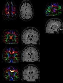
Magnetic Resonance Imaging
A dedicated 3 Tesla research MRI system is located in the Mellen Center on the main campus at Cleveland Clinic. This system is a Siemens TIM Trio MRI scanner operating under the Syngo operating system. This system has 32 receive channels.
The 3T MRI laboratory includes a Mallinckrodt power injector for precision bolus drug administration, equipment for ECG, pulse oximeter, and respiratory monitoring. A dual-bootable PC is situated adjacent to the scanner console and is capable of performing pulse sequence programming tasks, presentation of behavioral stimuli, and monitoring of physiologic information. A TTL synchronization signal is available from the scanner to synchronize stimulus presentation, behavioral responses, and physiologic information during scanning.
The 3 tesla Siemens Trio scanner is fully equipped with proton imaging and spectroscopy capability. It also has online capability to examine functional data superimposed on anatomical images in 3 dimension (Neuro 3D), which provides activation map and fieldmap information. MRI-compatible stimulus equipment includes Avotec, Inc.’s stereoscopic sound and stereoscopic visual presentation system, which can be used to present stimuli to subjects inside the MRI scanner.
The PC adjacent to the scanner console can monitor signals from a pair of fiber-optic gloves (Fifth Dimension Technologies, Inc., Irvine, CA). These are used to reliably monitor relative finger and hand position of subjects performing simple motor tasks inside the scanner. Signals from 15 fiber-optic strain gauges per hand are read-out every 25ms, giving a high temporal resolution measurement of the orientation of the joints of each finger and thumb.
Magnetic Resonance Resources
There are 10 MRI systems (three 3.0T, six 1.5T and two 1.0T) including three Siemens Avanto Tim multi-channel systems available on Cleveland Clinic’s Main Campus. Systems are equipped with the latest imaging coils and software capable state-of-the-art anatomic, physiologic and functional imaging.
Three 32-channel MRI systems (two Siemens Trio and one Philips Intera) are available. These machines have state-of-the-art gradient and coil systems for a wide variety of imaging. The systems are all equipped with multichannel receive chains allowing for parallel imaging. There are an additional 22 MRIs across the Cleveland Clinic that are primarily used for clinical imaging.
Nuclear Medicine Equipment
The Department of Nuclear Medicine in the Imaging Institute has a dedicated PET scanner (Siemens ECAT HR+) as well as a PET/CT (Siemens Biograph, LSO PET detectors, 16-row CT, contrast injector). PET/CT is list mode capable for dynamic or gated PET studies. Additionally the scanner has a laser patient positioning system. There are eight SPECT scanners, a radiopharmacy with 99mTc and 82Rb generators, workstations with clinical and research NM/PET analysis software, a nuclear cardiology core Lab, and extensive clinical databases for nuclear cardiology, PET/CT, cardiac PET.
Ultrasounds
There are 12 ultrasound systems that are capable of a full range of ultrasound imaging examinations. Systems have state-of-the-art transducers and software.
Facilities
Angiography Fluoroscopy Rooms
There are six angiography capable fluoroscopy rooms. Rooms include specialized equipment for peripheral, body, and neuroradiology angiography and interventional procedures. Specialized rotational angiography equipment is available for three-dimensional evaluation of aneurysms.
Imaging Archives
The Imaging Institute maintains a large imaging archive primarily for the storage of clinical images. The archive serves as both a primary image storage and retrieval site for many clinically-based trials throughout Cleveland Clinic. In other cases, the image archive accesses a backup storage device for raw imaging data which is stored in both raw and processed form on numerous individual databases at Cleveland Clinic.
The archive is a distributed system consisting of 20 separate imaging servers spread across nine separate Cleveland Clinic System Hospitals including Weston, Florida and Toronto, Canada. Ongoing imaging services produced more than two TB per week. The archive consists of 20 online SUN servers and two StorageTek Powderhorn tape systems that provide near-line access to images. Storage capacity across the enterprise is approximately 23TB of on-line storage. All images are archived to the near-line tape library. We have been archiving images near-line since 1994 with over 400 TB in storage. Storage facilities now allow for storage of 2 PB of data.
Laboratory
There is laboratory and office space for dedicated research personnel at the Mellen Center including 3 M.D. level staff, 5 Ph.D. level staff, 2 postdoctoral fellows and 2 master's level image analysis personnel. These labs contain general purpose test and measurement equipment (e.g. voltmeters, oscilloscopes, network analyzers). In addition, a state-of-the art 3 Tesla Siemens Trio MRI scanner is dedicated and staffed for biomedical research in a 200 sq. ft. imaging laboratory.
Mellen MR Imaging Center
The dedicated research 3 Tesla MR imaging facility at the Mellen Center is utilized for a wide range of MRI imaging studies. Facilities include laboratory space, fabrication facilities, high-end computing and data analysis equipment and software, as well as a state-of-the-art 3 Tesla Siemens Tim Trio MR system.
Post-Processing Lab
The laboratory provides advanced radiographic data manipulation services to both radiologists and clinicians of Cleveland Clinic and referring institutions, supporting the section's mission by maximizing diagnostic yield of CT and MR data sets and by optimizing staff radiologist workflow. The Lab processes neurovascular, coronary, abdominal and peripheral endovascular CT data, generating pre-rendered surface-shaded display and MIP data, as well as brain perfusion maps from both CT and MR data sets.
Hepatic and renal parenchymal volume measurements are performed, supporting the institutions busy and growing transplant services. Near term expansion of the number and scope of the Lab's services is expected to meet rapidly growing demand from both clinicians and radiologists.
Laboratory personnel and equipment are also available for data collection and analysis of research projects. Laboratory personnel are also available for research studies involving organization and archiving of large image data sets for research purposes. Facilities within the laboratory are available for research projects which require dedicated interpretation or analysis of image data sets.
