Cervical Spine

Your cervical spine consists of the first seven vertebrae in your spine. It provides support for the weight of your head, surrounds and protects your spinal cord, and allows for a wide range of head motions. Many conditions affect this area of your spine, including neck pain, arthritis, degenerative bone and disk disease, and stenosis. Many treatment options are available.

Overview

The Cervical Spine
The Cervical Spine

What is the cervical spine?

Your cervical spine — the neck area of your spine — consists of seven stacked bones called vertebrae. The first two vertebrae of your cervical spine are unique in shape and function. Your first vertebra (C1), also called the atlas, is a ring-shaped bone that begins at the base of your skull. It’s named after Atlas, of Greek mythology, who held the world on his shoulders. The atlas holds your head upright. Your second vertebra (C2), also called the axis, allows the atlas to pivot against it for the side-to-side “no” rotation of your head.

Your seven cervical vertebrae (C1 to C7) are connected at the back of the bone by a type of joint (called facet joints), which allow for the forward, backward and twisting motions of your neck.

Your cervical spine is also surrounded by muscles, nerves, tendons and ligaments. “Shock-absorbing” disks, called intervertebral disks, are positioned between each vertebra. Your spinal cord runs through the center of your entire spine. Your spinal cord sends and receives messages from your brain, which controls all aspects of your body’s functions.

Advertisement

Cleveland Clinic is a non-profit academic medical center. Advertising on our site helps support our mission. We do not endorse non-Cleveland Clinic products or services. Policy
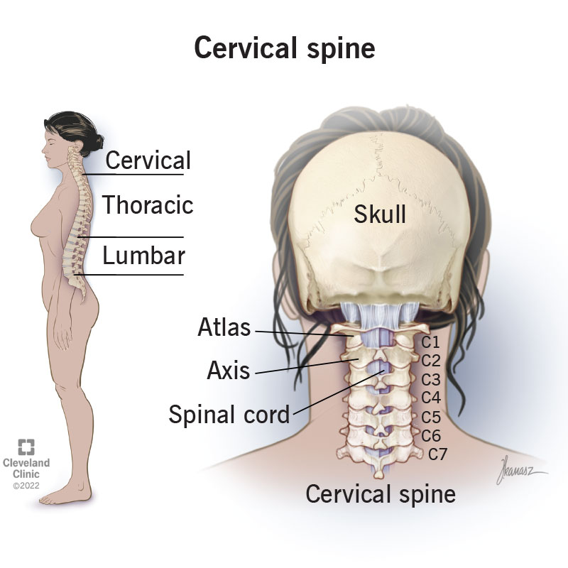
What does the cervical spine do?

Your cervical spine has several functions, including:

  • Protecting your spinal cord. The nerves of your spinal cord pass through a large hole (called the vertebral foramen) that passes through the center of all of your vertebrae — from the base of your skull through the cervical vertebrae, the thoracic (middle back) vertebrae and ending between the first and second lumbar (lower back) vertebrae. Taken together, all the stacked vertebrae of your spine form a protective central canal that protects your spinal cord.
  • Supporting your head and allowing movement. Your cervical spine supports the weight of your head (average weight of 10 to 13 pounds). It also allows your head and neck to tilt forward (flexion), backward (extension), turn from side to side (rotation) or bend to one side (ear-to-shoulder; lateral flexion).
  • Providing a safe passageway for vertebral arteries. Small holes in cervical spine vertebrae C1 to C6 provide a protective pathway for vertebral arteries to carry blood to your brain. This is the only section of vertebrae in the entire spine that contains holes in the bone to allow arteries to pass through.

What are the other muscles and soft tissues in the neck?

Other structures around or involving your cervical spine include the following:

Muscles supporting your cervical spine

The major muscles that attach to your cervical spine include:

  • Sternocleidomastoid. This muscle, one on each side of your neck, runs from behind your ear to the front of your neck. It attaches to your breast bone (sternum) and collarbone. This muscle allows you to rotate your head side-to-side and tilt your chin upward.
  • Trapezius. This pair of triangular muscles extend from the base of your skull down your cervical and thoracic spine and out to your shoulder blade. They help tilt your head upward/move your neck backward, rotate your head right or left or lift your shoulder blade.
  • Levator scapulae. This muscle attaches to your first four cervical vertebrae and the top of your shoulder blade (scapula). It helps lift your shoulder blade, bend your head to the side and rotate your head.
  • Erector spinae. Several muscles make up this muscle group. In your cervical spine area, these muscles help with posture, neck rotation and backward neck extension.
  • Deep cervical flexors. These muscles run down the front of your cervical spine. They allow you to flex your neck forward neck and help keep your cervical spine stable.
  • Suboccipital muscles. These four pairs of muscles connect the top of your cervical spine with the base of your skull. They allow you to extend and rotate your head.

Ligaments of your cervical spine

Ligaments in your cervical spine connect bone to bone to help to keep your cervical spine stable. Three major cervical spine ligaments are:

  • Anterior longitudinal ligament. This ligament extends from the base of your skull, down the front of the cervical vertebra. It stretches to resist backward neck motion.
  • Posterior longitudinal ligament. This ligament starts at C2 and extends down the back of your cervical vertebrae. It stretches to resist forward neck motion.
  • Ligamentum flava. These ligaments line the backside of the inside opening of each vertebra where your spinal cord passes. These ligaments cover and protect your spinal cord from behind.

Disks in the cervical spine

Cervical disks are the “shock absorber cushions” that sit between each vertebra. A total of six disks are positioned between the seven cervical vertebrae (one between two vertebrae). In addition to cushioning against stresses placed on your neck, the disks allow you to flex and rotate your head more easily during activity.

Nerves in the cervical spine

Eight pairs of spinal nerves exit through small openings (foramen) between every pair of vertebrae in your cervical spine. They’re labeled C1 through C8. They stimulate muscle movement in your neck, shoulder, arm and hand, and provide sensation.

  • Cervical nerves C1, C2 and C3 control your forward, backward and side head and neck movements. The C2 nerve provides sensation to the upper area of your head; C3 gives sensation to the side of your face and back of your head.
  • Cervical nerve 4 controls your upward shoulder motion and is one of the nerves that controls your diaphragm (muscle at the bottom of your rib cage that helps you breathe). C4 provides sensation for parts of your neck, shoulders and upper arms.
  • Cervical nerve 5 controls the deltoid muscles of your shoulders and your biceps. C5 provides sensation to the upper part of your upper arm down to your elbow.
  • Cervical nerve 6 controls the extensor muscles of your wrist and is involved in the control of your biceps. C6 provides sensation to the thumb side of your forearm and hand.
  • Cervical nerve 7 controls your triceps and wrist extensor muscles. C7 provides sensation to the back of your arm into your middle finger.
  • Cervical nerve 8 controls your hands and gives sensation to the pinky side of your hand and forearm.

Spinal cord

Your spinal cord is a bundle of nerve tissue that extends from the lower part of your brain to your body. It carries messages between your brain and the muscles mentioned above.

Advertisement

Additional Common Questions

What diseases and disorders affect your cervical spine?

Many diseases and conditions result from problems in the cervical spine and the surrounding soft tissues and nerves. These include:

  • Cervical radiculopathy. This condition arises when a cervical nerve is pinched by cervical vertebrae. You may experience tingling, numbness, weakness and pain. Symptoms may remain local or can spread to your entire arm, hand and fingers. Cervical radiculopathy is also called a pinched nerve or cervical nerve compression.
  • Neck pain. Neck pain is a common symptom of many different injuries and medical conditions. Common causes include degenerative conditions (osteoarthritis, spinal stenosis, herniated disk, pinched nerve), whiplash, mental stress, physical strain, poor posture, growths (tumors, cysts, bone spurs), meningitis, rheumatoid arthritis and cancer.
  • Cervical degenerative disk disease. Cervical degenerative disk disease occurs when the disks in your cervical spine wear down.
  • Herniated disk. This condition is a tear or leak to the disks that provide a cushion between vertebrae. Intervertebral disks allow you to bend and move with ease.
  • Bone spurs in your cervical spine (cervical osteophytes). Bone spurs are growths that occur on any of the seven vertebrae in your cervical spine.
  • Cervical spondylosis. Cervical spondylosis, also called arthritis of the neck, is the age-related slow degeneration of your disks and joints in your cervical spine
  • Cervical spinal cord injury. A cervical spinal cord injury is an injury to your cervical vertebrae. Most spinal cord injuries are the result of a sudden, traumatic blow to the vertebrae.
  • Cervical spinal fracture. A fracture to the bones of your spine can result from compression (often from minor trauma in a person with osteoporosis) or be a burst fracture (vertebra that’s crushed in all directions) or a fracture-dislocation (mostly from vehicle accidents or falls from heights).
  • Cervical spinal cord compression (cervical spondylotic myelopathy). This is a condition in which there’s pressure on your spinal cord in the cervical area of your spine. One of the most common causes is wear and tear on the bones of your spine, a condition called osteoarthritis.
  • Cervical stenosis. This condition occurs when your spinal canal in the cervical spine area narrows. Less space within your cervical spine reduces the amount of space available for your spinal cord and nerves that branch off the spinal cord. A tightened space can cause your spinal cord or nerves to become irritated, compressed or pinched.
  • Cervical spinal tumor and cancer. Tumors are abnormal growths of tissue inside your spinal column. They can either be noncancerous (benign) or cancerous (malignant).
  • Meningitis. Meningitis is an infection of the meninges. The meninges are a protective lining around your brain and spinal cord.
  • Osteomyelitis. Osteomyelitis is a bacterial or fungal infection of the bone, in this case, the vertebrae of your spine. If left untreated, it can lead to the death of vertebrae.

How are diseases and conditions of the cervical spine diagnosed?

First, your healthcare provider will gather your medical and medication history, ask you about your symptoms, perform a physical exam and order tests and imaging studies.

Tests and imaging may include:

  • Computed tomography (CT) scan. This scan uses X-rays and computers to produce images that are very thin “slices” of the area under examination. A CT scan can show the shape and size of your spinal canal, its contents and the bone around it. It helps diagnose bone spurs, osteophytes, bone fusion and bone destruction from infection or tumor.
  • Magnetic resonance imaging (MRI). This test uses a large magnet, radio waves and a computer to produce detailed images. This scan can reveal problems with your spinal cord and nerves exiting the spinal column, spinal degeneration, disk herniation, infections and tumors.
  • X-rays. X-rays create pictures of your bones and soft tissues, using a small amount of radiation. X-rays can show fractures, disk problems, spinal alignment problems and the presence of arthritis.
  • Electromyogram (EMG) and nerve conduction studies. An EMG helps evaluate the health and function of nerves and muscles. A nerve conduction study measures how fast an electrical impulse moves through your nerve. These tests help determine ongoing nerve damage and the site of nerve compression.
  • Myelogram. This imaging test examines the relationship between your vertebrae and disks, outlines the spinal cord and nerves exiting your spinal column. It shows if such possible things as a tumor, bone spurs or herniated disk are pressing against your spinal cord, nerves or nerve roots and causing pain, numbness or weakness.
  • X-rays. X-rays create pictures of your bones and soft tissues, using a small amount of radiation. X-rays can show fractures, disk problems, spinal alignment problems and the presence of arthritis.
Advertisement

How are cervical spine health issues treated?

Both nonsurgical treatment options and surgery are available to treat many of the conditions that affect the cervical spine. The choice depends on the cause of the cervical spine issue and its severity.

What are the nonsurgical treatment options for cervical spine conditions?

Your healthcare provider may first recommend less invasive approaches for neck pain that aren’t caused by trauma or a tumor. Some common nonsurgical treatment options include:

  • Rest.
  • Ice or heat.
  • A soft cervical collar. A collar helps support and immobilize your neck.
  • Avoiding strenuous or aggravating physical activity.
  • Physical therapy.
  • Medications, including muscle relaxants, pain relievers (such as acetaminophen) and anti-inflammatories (such as ibuprofen and naproxen).
  • Steroid injections. Two specific types of steroid injections may be considered for neck and/or arm pain. A cervical epidural block is a procedure in which the steroid is injected into the epidural space (the space next to the covering of youe spinal cord). Cervical facet joint block is a procedure in which the steroid is injected into the capsule (connective tissue covering) of the facet joint (the small joint at the top and bottom of each vertebra that connects the vertebrae to permit motion).
  • Medical branch block and radiofrequency ablation. This procedure is considered in some cases of chronic neck pain. First, a local anesthetic is injected into the nerve that supplies the facet joint of the vertebrae. If your pain is relieved, the next step is to make pain relief permanent. This is done by damaging your nerve with a technique called radiofrequency ablation. Pain relief lasts for months. If your nerve regenerates, the pain can return.

How do I know if I’m a candidate for cervical spine surgery?

You may be a candidate for cervical spine surgery if:

  • Other treatments aren’t helping.
  • Symptoms involving your spine, arms and/or legs are worsening.
  • You’re healthy enough to have surgery.

What surgical treatment options are available for cervical spine conditions?

Common surgical approaches include:

Cervical spinal decompression surgery

Cervical spinal decompression surgery is a general term that refers to various procedures used to relieve symptoms caused by pressure, or compression, on your spinal cord or nerve roots. Nerve roots are the first segment of a nerve that leaves your spinal cord through the small hollows between the vertebrae. Common surgical techniques for decompression include:

  • Cervical diskectomy. In this procedure, your surgeon removes a portion of a disk to relieve pressure on the nearby nerve roots.
  • Cervical laminotomy or laminectomy. In these procedures, your surgeon removes a small part of the bony arches of the spinal canal, called the lamina. Only a small section of the lamina is removed in a laminotomy. The entire lamina is removed in a laminectomy along with any bone spurs, disk material and thickened ligament if needed. Removing the lamina increases the size of the spinal canal, which relieves pressure.
  • Cervical foraminotomy or foraminectomy. Both of these procedures are performed to expand the openings for the nerve roots to exit your spinal cord by removing some bone in that area. In a foraminectomy, a large amount of bone is removed.
  • Cervical corpectomy. In this surgery, your surgeon removes the body of the vertebra (the large front portion of the vertebra), as well as the disk to relieve pressure on the spinal cord. In some cases, this is followed by fusion of the vertebrae (permanently connecting two or more vertebrae) to keep your cervical spine stable.

Cervical disk replacement surgery

Cervical disk replacement surgery involves removing a diseased cervical disk and replacing it with an artificial disk. The most common reason for this procedure is cervical disk degeneration.

Cervical spinal fusion

Cervical spinal fusion is surgery that permanently connects to one or more cervical vertebrae. The surgery eliminates the motion between vertebrae.

Functional electrical stimulation for spinal cord injury

Functional electrical stimulation for spinal cord injury. This procedure uses small electrical impulses to activate specific muscles and nerves to restore function to your upper body muscles controlled by cervical nerves.

Is having minimally invasive cervical spine surgery a possibility?

Speak with your surgeon. In many cases, minimally invasive spine surgery is an option. Compared to the one large incision through your skin with traditional open surgery, minimally invasive surgery is performed through one or more smaller incisions. Working through smaller incisions causes much less damage to muscles and soft tissues than a single long incision.

A note from Cleveland Clinic

Your cervical spine is the neck region of your spinal column or backbone. It consists of your first seven bones (C1-C7). Other structures in or around your cervical spine are your intervertebral disks, spinal cord and nerves, muscles, tendons and ligaments. Your cervical spine supports the weight of your head and allows a wide range of head movement. Its circular surround of bone also protects your spinal cord. Many diseases and disorders can affect your cervical spine. Fortunately, many nonsurgical and surgical options can treat these conditions.

Medically Reviewed

Last reviewed by a Cleveland Clinic medical professional on 01/18/2022.

Learn more about our editorial process.

Ad
Appointments 866.588.2264