Breast MRI

A breast MRI is an imaging test that produces very clear pictures of breast tissue. Healthcare providers mainly use it to evaluate known breast cancer. But they also use it in addition to mammography and ultrasound to screen for and diagnose breast cancer and other abnormalities.

Overview

Breast MRI Questions

What is a breast MRI?

Breast MRI (magnetic resonance imaging) is a type of imaging study that uses a large magnet, radio waves and a computer to produce detailed images of breast tissue. Unlike mammograms, a breast MRI doesn’t use X-rays (radiation).

Some of the most common reasons to perform a breast MRI include:

  • To screen people who are at high risk for developing breast cancer because they have a strong family history, carry genes that increase their risk and/or have dense breast tissue. MRI is important because it may detect cancers that mammograms or ultrasounds can’t detect.
  • To evaluate the extent of the cancer in people who have a new diagnosis of breast cancer.
  • To evaluate for silicone breast implant ruptures.

Depending on the reason for a breast MRI, it may or may not require injection of contrast dye (called gadolinium). For many breast MRIs, a technologist or nurse will place an IV in your arm so they can inject contrast dye during the test. This helps a radiologist see normal and abnormal structures in your breast more clearly.
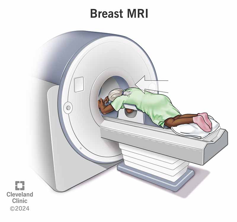
 This dye is different from the one used during CT scans.

Advertisement

Cleveland Clinic is a non-profit academic medical center. Advertising on our site helps support our mission. We do not endorse non-Cleveland Clinic products or services. Policy

What is the difference between a breast MRI and a mammogram?

Healthcare providers use mammograms and breast MRIs (and ultrasounds) to help detect breast cancer and other abnormalities.

However, mammograms are usually the first option for screening and diagnosis of breast cancer, particularly as mammograms are the only imaging test able to reliably identify microcalcifications — an early presenting sign of some breast cancers.

There are also differences in how the two imaging tests work.

Mammogram
Uses X-rays.
Breast MRI
Doesn’t use X-rays.
Compresses (squeezes) your breasts.
Breast MRI
Doesn’t compress your breasts.
Doesn’t use contrast dye (no IV necessary).
Breast MRI
Usually requires contrast dye (an IV is necessary).
Images acquired over two to three minutes.
Breast MRI
Images acquired over 17 to 20 minutes.

Why would my healthcare provider order a breast MRI?

Your healthcare provider may recommend a breast MRI for several reasons, including:

  • As part of breast cancer screening in addition to mammography if you’re at high risk for breast cancer (usually because of strong family history or genetic mutations, like BRCA1 or BRCA2).
  • To gather more information after you’ve been diagnosed with breast cancer.
  • To further evaluate breast abnormalities that are difficult to assess with mammography and breast ultrasound.
  • To evaluate breast implants. MRI is the best imaging test to see if silicone breast implants have ruptured.

Breast MRI and breast cancer

Providers often recommend this test to provide more detail after diagnosing breast cancer. It can:

  • Measure the extent of the cancer and see if it extends into your nipple, skin or underlying chest wall muscle.
  • Look for other cancer or abnormal tissue in your breast.
  • Check if you have any large lymph nodes in your armpit, which can be a sign that the cancer has spread (metastasized) to that area.
  • Monitor the progress of breast cancer treatment, especially chemotherapy to shrink the tumor before surgery (neoadjuvant chemotherapy).

Breast MRIs may also be a helpful test to further evaluate lumpectomy sites in the years following breast cancer treatment. This is because scarring and recurrent breast cancer can look the same on mammography and ultrasound.

Does a breast MRI always show cancer?

An MRI can find some small breast tumors that a mammogram might miss, but it can also produce false-positive and false-negative results. For example, sometimes, a benign (noncancerous) piece of tissue in your breast can show up as a bright spot on the image, much like breast cancer. This would be a false-positive result. In any case, if a radiologist sees something in your breast tissue that doesn’t look normal, they may order additional tests such as a breast ultrasound or a biopsy to determine exactly what the finding is.

Alternatively, not all cancers will always show up on MRI that radiologists may be able to better see on other imaging tests, like a mammogram. This would be a false-negative result. This is one reason why the American Cancer Society and the U.S. Preventative Services Task Force don’t recommend breast MRI as a screening test by itself.

Advertisement

Test Details

A person lying on their stomach on an exam table going head-first into the opening of an MRI machine, shaped like a donut.
A breast MRI uses a large magnet, radio waves and a computer to produce detailed images of breast tissue.

Who performs a breast MRI?

A radiologist or radiology technologist performs a breast MRI.

A radiology technologist is a medical professional who’s specially trained and certified to perform MRI scans, but they don’t interpret the results or give medical opinions or advice.

A radiologist is a medical doctor who performs and interprets imaging tests to make a diagnosis. They offer medical advice based on the findings.

How do I prepare for a breast MRI?

Before your breast MRI, it’s important to tell your provider if you:

  • Are pregnant or breastfeeding/chestfeeding.
  • Are unable to lie on your stomach for 30 to 60 minutes.
  • Have had an allergic reaction to a contrast dye in the past.
  • Have claustrophobia (fear of closed or narrow spaces). You may be able to take a sedative to help you relax for the exam.

The magnetic resonance imaging (MRI) scanner uses a strong magnet and radio wave signals that can cause heating or possible movement of some metal objects in your body. This could result in health and safety issues. It could also cause some implanted electronic medical devices to malfunction.

If you have metal-containing objects or implanted medical devices in your body, your healthcare team needs to know about them before your exam. Please tell your provider and MRI technologist if you have any of the following:

  • Heart pacemaker/defibrillator.
  • Electronic/implanted stimulators or devices, such as a vagus nerve stimulator.
  • Cochlear implant or other ear implants.
  • Aneurysm clips and coils.
  • Stents not located in your heart.
  • Filters, such as blood clot filters.
  • Metal fragments in your body or eye, such as bullets, metal shrapnel or shavings, etc.

You won’t be able to wear the following devices during your MRI:

Talk to the provider you see for these devices (such as your endocrinologist) to learn how you should go about the MRI.

Advertisement

What should I expect during a breast MRI?

During a breast MRI, you can expect the following:

  1. You’ll change into a medical robe.
  2. If you’re getting the MRI for anything other than examining a breast implant, a provider will insert an IV line into a vein in your arm to inject the contrast dye during the exam.
  3. You’ll lie face down on a table that has openings for your breasts. The technologist will help you get into the correct position. Let them know if you’re uncomfortable.
  4. The technologist will move the table into the magnet of the MRI machine. They’ll perform the imaging test with a computer while watching you through a window in a different room. You’ll be able to talk to the technologist via an intercom if needed.
  5. While the machine is taking images, it’s important to remain as still as possible to ensure the images aren’t blurry. The MRI machine will make a loud banging noise throughout the entire exam. This means the machine is working properly. The technologist will give you earplugs to help you through the exam.
  6. If your MRI test requires a contrast dye, the technologist will inject it after an initial series of scans. They’ll take more images during or following the injection.
  7. Once the test is done, the technologist may ask you to wait while the radiologist checks the images in case they need more.
  8. Once they’ve taken enough pictures, the technologist will remove your IV line and place a bandage over the site. The test is now over.

Many people are nervous about an MRI scan because they’re afraid of being enclosed in a tight space. It’s true that the older machines were narrow with very little head-to-ceiling space. But newer machines are larger and have significantly improved in terms of comfort. For example, newer MRI machines are open at both ends. They also have wider openings, shorter total lengths, more head-to-ceiling space and are fully ventilated (a fan will blow a gentle stream of air on you).

If you’re worried you may be too anxious during the procedure, talk to your provider. You may be able to take a sedative before the procedure so you’re more relaxed.

What should I expect after my breast MRI?

Generally, you can resume your usual activities after a breast MRI.

However, if your exam was an MRI breast biopsy, follow the at-home instructions your provider gives you. If you took a sedative for your exam, you’ll need someone else to drive you home.

How long does a breast MRI take?

Although many facilities plan for 40 to 60 minutes for a breast MRI, the scan itself only lasts up to 20 minutes. The extra time is used for screening questionnaires, IV placement and proper positioning for the exam.

What are the risks of a breast MRI?

There’s very little risk to getting a breast MRI as long as you and your technologist follow the safety guidelines.

There’s a chance you might have an allergic reaction to the contrast dye (gadolinium), but this is very rare. Reactions are usually mild, and your healthcare team will quickly treat it if it happens.

It’s important to tell your provider and technologist if you’re pregnant or breastfeeding (chestfeeding).

Care at Cleveland Clinic

Results and Follow-Up

What type of results do you get from a breast MRI?

Depending on whether you got a breast MRI for screening purposes, diagnostic purposes or to evaluate known cancer, your MRI report may look different. In any case, your provider who ordered the breast MRI will discuss your results with you.

Radiologists all use the same standardized system to describe screening and diagnostic breast imaging (including mammogram, ultrasound and MRI) results. This system is called the Breast Imaging Reporting and Data System (BI-RADS). This system categorizes results on a scale of 0 through 6.

BI-RADS category
0
Definition
Incomplete.
Explanation
This result means the radiologist may have seen a possible abnormal area, but they need further images to evaluate it, such as a diagnostic mammogram or an ultrasound. This result may also mean that the radiologist wants to compare your most recent breast MRI with older ones to see if there’ve been changes in the area over time.
1
Definition
Negative.
Explanation
This result means the radiologist didn’t find a significant abnormality on the images.
2
Definition
Benign (noncancerous) finding.
Explanation
This result means that the radiologist found a benign (noncancerous) area in your breast, such as benign cysts, lymph nodes or fibroadenomas. The radiologist records this finding to help when comparing it to future breast imaging tests.
3
Definition
Probably benign finding.
Explanation
The findings in this category have a greater than 98% chance of being benign (noncancerous). But as it’s not proven to be benign, the radiologist wants to monitor it to be sure it doesn’t change over time. You’ll likely need additional imaging (such as MRI) in six months.
4
Definition
Suspicious abnormality.
Explanation
This result means a finding(s) is not normal and has suspicious features suggesting it could be cancer. The radiologist will recommend a breast biopsy to get more information. The findings in this category can have a 2% to 95% chance of being a cancer.
5
Definition
Highly suggestive of malignancy.
Explanation
The term “malignancy” refers to the presence of cancerous cells. This result means the findings look like cancer and have at least a 95% chance of being cancer. The radiologist will strongly recommend a breast biopsy of these findings.
6
Definition
Known biopsy-proven malignancy.
Explanation
Radiologists use this result for findings on a mammogram or MRI that’ve previously been biopsied and are a known cancer. Healthcare providers use MRIs in this way to see the extent of the cancer and/or how well it’s responding to treatment.

When should I know the results of my breast MRI?

The results of your MRI will be available in your electronic medical records account (if you have one) and should be available to the provider who ordered the test within one to two business days of your exam. Your provider will discuss the results with you.

A note from Cleveland Clinic

A breast MRI is a helpful tool for screening, diagnosing and evaluating breast cancer and other abnormalities. If you need a breast MRI and are worried about the procedure or have questions about it, don’t be afraid to ask your healthcare provider. They’re available to help and support you.

Medically Reviewed

Last reviewed by a Cleveland Clinic medical professional on 12/13/2022.

Learn more about our editorial process.

Ad
Appointment Center 216.445.7050