An intravenous pyelogram is a type of X-ray that creates images of your urinary system. Healthcare providers inject dye into a vein, which an X-ray picks up. In most cases, healthcare providers no longer use this test.
Advertisement
Cleveland Clinic is a non-profit academic medical center. Advertising on our site helps support our mission. We do not endorse non-Cleveland Clinic products or services. Policy
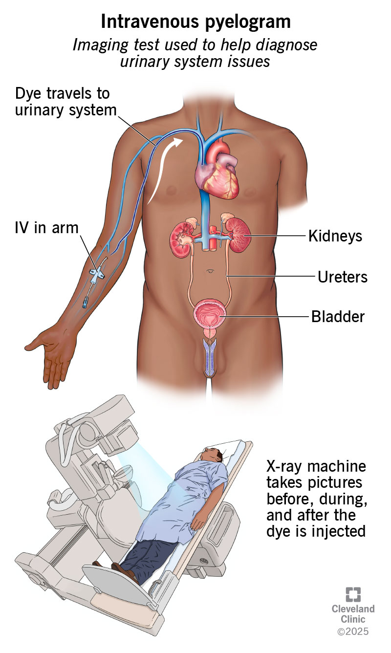
Image content: This image is available to view online.
View image online (https://my.clevelandclinic.org/-/scassets/images/org/health/articles/intravenous-pyelogram)
An intravenous pyelogram (in-truh-VEE-nuhs pahy-EL-uh-gram) is a type of imaging test. It shows how your urinary system handles pee. It can help diagnose problems in your kidneys, ureters and urinary bladder.
Advertisement
Cleveland Clinic is a non-profit academic medical center. Advertising on our site helps support our mission. We do not endorse non-Cleveland Clinic products or services. Policy
During the test, healthcare providers inject a contrast dye into a vein. The dye travels through your blood to your urinary system. X-rays pick up the dye, which makes the parts of your urinary system look white in the images.
Other names for an intravenous pyelogram include:
Today, intravenous pyelogram isn’t common. Healthcare providers are more likely to recommend other imaging tests, like:
Healthcare providers rarely perform IVP. But in the past, they may have recommended IVP to diagnose:
Providers sometimes recommend IVP to help diagnose:
Healthcare providers will review your medical history before an IVP. Be sure to tell them if you:
Your provider will also give you specific instructions on eating or drinking before an IVP. And they may recommend taking a laxative so you poop. Empty bowels can make it easier for your provider to see any problems in your images.
Advertisement
No. You should pee right before the test. This makes it easier for healthcare providers to see the contrast dye in your urinary system.
A radiologist or radiologic technologist will perform an intravenous pyelogram. Radiologists are medical doctors who specialize in diagnosing conditions using imaging tests. Radiologic technologists aren’t doctors. But they can perform some tests, including IVPs.
In general, your healthcare provider will:
An IVP usually takes about an hour, and you should lie still as they take the images. But it may take longer if your kidneys function slowly.
Afterward, your healthcare provider will remove the peripheral IV from your arm. They’ll place a bandage over the area where the needle went in. They’ll also encourage you to drink lots of fluids. Drinking fluids, especially water, helps flush the dye out of your system. Afterward, you can continue your regular activities.
You’ll likely feel some symptoms after your healthcare provider injects the dye. These symptoms usually don’t last long. They may include:
In some cases, you may feel nauseous or throw up. Your face may also feel flushed.
Severe allergic reactions to contrast dye are rare. But they’re possible. They may include:
Healthcare providers may not recommend IVP if you have:
It depends on the conditions that affect your urinary system. An IVP may reveal:
A radiologist will look for anything unexpected in the images. They’ll then report to the healthcare provider who recommended an IVP. Your provider will contact you to explain the results. They’ll also discuss any next steps in your treatment.
Advertisement
You should expect to get your results within a few days.
If your results are abnormal, your healthcare provider may recommend more tests. Tests may include:
They may also recommend treatment.
After your IVP, call your healthcare provider if you have any symptoms that may point to problems with your urinary system. These may include:
Get to the nearest emergency room if you show signs of a severe allergic reaction to contrast dye.
There’s no difference between an intravenous pyelogram and an intravenous urogram (IVU). They’re different names for the same test.
An intravenous pyelogram can help identify problems in your urinary system. But these days, there are other imaging tests that are more comfortable and take more detailed images. If you need an IVP, you may feel better knowing it’s generally safe with low chances of complications. You can also return to your usual activities right after the test. If you have any questions or concerns, reach out to a healthcare provider.
Advertisement


Sign up for our Health Essentials emails for expert guidance on nutrition, fitness, sleep, skin care and more.
Learn more about the Health Library and our editorial process.
Cleveland Clinic’s health articles are based on evidence-backed information and review by medical professionals to ensure accuracy, reliability and up-to-date clinical standards.
Cleveland Clinic’s health articles are based on evidence-backed information and review by medical professionals to ensure accuracy, reliability and up-to-date clinical standards.
If you have a condition that’s affecting your urinary system, you want expert advice. At Cleveland Clinic, we’ll work to create a treatment plan that’s right for you.
