Your inner ear is the innermost part of your hearing system and home to your vestibular (balance) system. It contains the cochlea, which helps you hear, and the semicircular canals and otolith organs that help you balance. You can protect your inner ear by avoiding things, like loud noises, that can damage the sensitive structures inside.
Advertisement
Cleveland Clinic is a non-profit academic medical center. Advertising on our site helps support our mission. We do not endorse non-Cleveland Clinic products or services. Policy
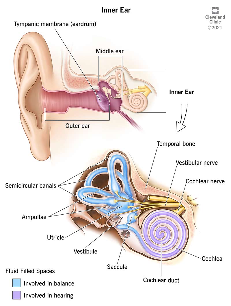
Image content: This image is available to view online.
View image online (https://my.clevelandclinic.org/-/scassets/Images/org/health/articles/24340-inner-ear)
What we think of as the “ear” is actually a three-part structure. The outer ear is the part you see and your ear canal. The middle ear is a box-shaped area behind your tympanic membrane (eardrum) that includes the three smallest bones in your body. And the inner ear is just beyond the middle ear, in a small hole in the temporal bones that help make up the sides of your skull.
Advertisement
Cleveland Clinic is a non-profit academic medical center. Advertising on our site helps support our mission. We do not endorse non-Cleveland Clinic products or services. Policy
Although the structures within your inner ear are tiny and tucked away, they play a huge role in your outward experience of the world — both the sounds you hear and feelings of equilibrium within your body.
Your inner ear has two jobs:
Your inner ear is the last stop that sound waves make in a carefully orchestrated journey that starts from your outer ear. These waves travel from your outer ear through your middle ear to your inner ear. Structures within your inner ear convert the sound waves into electrical energy. Your hearing (auditory) nerve delivers the energy to your brain as sound, making it possible for you to hear.
At the same time, your inner ear monitors your movements. It alerts your brain to changes in your position, so your brain can let your body know what to do to stay balanced.
Your inner ear includes two parts: the cochlea (which supports your hearing) and the vestibular system organs (which support your balance).
Your cochlea is a snail-shaped organ within your inner ear that helps you hear. It’s filled with fluid that moves in response to sound waves and is split into three tubes by two thin membranes. One of these membranes — the basilar membrane — is like an elastic wall, on top of which sits the organ of Corti. The organ of Corti contains tiny hair cells with stereocilia at the ends. Stereocilia are delicate, hair-like projections that react to cochlea fluid movement.
Advertisement
Here’s how your cochlea turns sound waves into sounds:
The structures inside your inner ear that help you balance are part of your vestibular system. They include three fluid-filled semicircular canals and two otolith organs (the saccule and utricle).
Here’s what the parts are and what they do:
Movement of hair cells triggers an electrical impulse that travels from your vestibulocochlear nerve, or the 8th cranial nerve, to your brain. Your brain perceives the electrical signal as information about your balance.
Your hearing and your sense of balance rely on a healthy inner ear. The most common inner ear conditions and disorders that can get in the way of this include:
Advertisement
Other conditions that can cause inner ear problems include:
Signs of an inner ear problem include:
You may experience other symptoms depending on the specific inner ear condition.
Healthcare providers use several tests to check the inner ear and diagnose inner ear problems, including:
Depending on your diagnosis, treatments may involve managing or curing your condition. Common treatments include:
Advertisement
There are several ways to protect your inner ear from hearing loss. Here are some important ones:
Your inner ear is a complicated, delicate and essential part of your body. Every minute of every day, your inner ear turns sound waves into sounds that keep you safe and enrich your life. It also helps you stay balanced. Hearing loss and balance problems can come without warning and interfere with how you’re able to use the sensory information associated with your inner ear. Without treatment, inner ear problems and can worsen over time. Talk to your healthcare provider as soon as you suspect something might be wrong with your inner ear.
Advertisement


Sign up for our Health Essentials emails for expert guidance on nutrition, fitness, sleep, skin care and more.
Learn more about the Health Library and our editorial process.
Cleveland Clinic’s health articles are based on evidence-backed information and review by medical professionals to ensure accuracy, reliability and up-to-date clinical standards.
Cleveland Clinic’s health articles are based on evidence-backed information and review by medical professionals to ensure accuracy, reliability and up-to-date clinical standards.
Hearing is an important part of your everyday life. Hearing loss can impact your life in so many ways. Cleveland Clinic experts can help you hear clearly again.
