



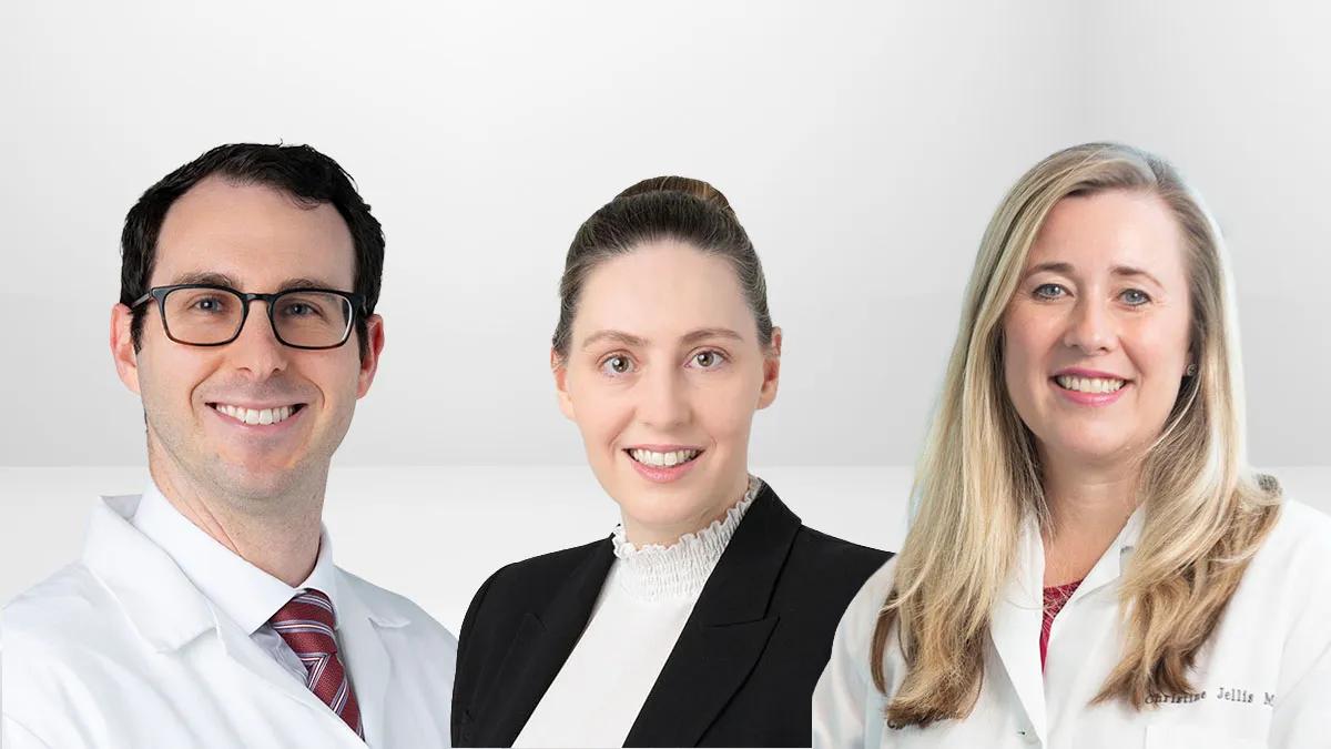
Talking Tall Rounds® Cardiac Sarcoidosis: Systematic Team Approach to Diagnosis & Management
In this excerpt from Tall Rounds®, Cleveland Clinic experts discuss imaging modalities that aid the diagnosis of cardiac sarcoidosis. View other Tall Rounds programs.
Meet the presenters:
Amanda Vest, MBBS
Christine Jellis, MD
Bryan Abadie, MD
Looking to refer a patient? Please reach out to our Physician Referral team Mon. - Fri., 8 a.m. - 5 p.m. (ET) at 855.751.2469.
Advertisement
Cleveland Clinic is a non-profit academic medical center. Advertising on our site helps support our mission. We do not endorse non-Cleveland Clinic products or services. Policy
Recent Episodes
Evolving Strategies in Aortic Arch Repair
Eric Roselli, MD, and Patrick Vargo, MD, discuss how advances in open, hybrid and endovascular techniques are changing the treatment of aort…
Management of Complex Aortic Valve Endocarditis
Milind Desai, MD, and Per Wierup, MD, discuss the evaluation and management of high-risk aortic valve endocarditis, including indications fo…
Lepodisiran and the Emerging Role of Oxidized Phospholipids in Lipoprotein(a) Risk
Luke Laffin, MD, speaks with Steven Nissen, MD, about findings from the ALPACA trial evaluating lepodisiran and its effects on oxidized phos…
Video content: This video is available to watch online.
View video online (https://cdnapisec.kaltura.com/p/2207941/sp/220794100/playManifest/entryId/1_e799pgn2/flavorId/1_5f3sgelj/format/url/protocol/https/a.mp4)
Transcript
Announcer:
Welcome to Cardiac Consult, brought to you by the Sydell and Arnold Miller Family Heart, Vascular & Thoracic Institute at Cleveland Clinic. This podcast will explore the latest innovations, medical and surgical treatments, diagnostic testing, research, technology, and practice improvements.
Advertisement
Cleveland Clinic is a non-profit academic medical center. Advertising on our site helps support our mission. We do not endorse non-Cleveland Clinic products or services. Policy
Dr. Amanda Vest, MBBS:
Well, good morning, everybody. Welcome to today's Tall Rounds. We're delighted to have a topic today of cardiac sarcoidosis. As you'll see from our presenters gathered here today, this is a highly multidisciplinary, multi-specialty condition. We all participate in a multidisciplinary cardiac sarcoidosis case presentation and discussion every two weeks, which really allows us to learn from each other and bring together all the complementary skills within the group and try to figure out both diagnosis, which can sometimes be quite a conundrum in this patient population, as well as optimal treatments. I'm delighted that several of my colleagues are going to be sharing their expertise with us today as to how do we think through these cases, especially patients with potentially isolated cardiac sarcoidosis, but also when cardiac sarcoidosis is part of a multi-system disease. Not only has the pathophysiological understanding moved forward, but our ability to diagnose cardiac sarcoid has nuanced, and certainly MRI is a big part of that.
So, Dr. Jellis will take us through echo and MRI.
Dr. Christine Jellis, MD:
Thank you, Dr. Vest. As we've heard, tissue can be king, but it's often difficult to get a myocardial biopsy. We rely heavily on our advanced cardiac imaging to help us find a source of truth. As you'll see here though, sometimes it feels like we're truly in an epidemic of sarcoidosis, but I would put to you that this is really the evolution of advanced imaging. As you'll see, over time with these studies, the predicted prevalence of cardiac sarcoidosis has certainly seemed to rise, but is this just because we're making the invisible visible by our advanced testing?
Advertisement
Echocardiography remains the mainstay of the tools that we use in our imaging lab. Many of you will be familiar with the commonly known 2D manifestations that we can see on our echocardiograms. In particular, the regional wall motion and abnormalities, these focal aneurysms in non-coronary distributions, along with left ventricular systolic, diastolic dysfunction and other features like pericardial effusions.
We typically know that the basal inferior septum is the largest area where we see macroscopic involvement, largely explaining why many of these patients have a heart block as part of their presentation. But we can also see that there are just other features that are a little bit characteristic of sarcoid, with the caveat that sarcoidosis can certainly masquerade as many things. However, as you'll see, echocardiography, while helpful, has a relatively low sensitivity. We really need to overlay this with more advanced imaging.
Initially, we turn to a global longitudinal strain, and you can see here that in patients with extracardiac sarcoidosis, a cut point of a strain that's less negative than minus 17.3 can be used as a determinant of someone that's likely to have cardiac sarcoidosis in the setting of their extracardiac disease. That can certainly be helpful in guiding us along with other parameters such as ejection fraction and the E/e’ (E-on-e prime) ratio.
You can see here the bullseye plots there, normal being completely red and abnormal where you can see the light pink come in, which gives us a rather sort of global outlook on where we think the strain may be regionally reduced. That reduced strain does correspond to patients who are likely to have positive evidence of LGE (late gadolinium enhancement) on an MRI, and therefore can improve our ability to detect sarcoidosis incrementally on top of our standard echo imaging. You can see here some nice examples where in the bullseye you can see the reduced pink area of strain, obviously corresponding to this focal area of thinning and aneurysmal deformation in the inferoseptum, despite the ejection fraction being relatively preserved.
As that brings us to cardiac MRI, I highlight here with the red circles, the segments where typically we do see more involvement with evidence of cardiac sarcoidosis. However, as I also alluded to, sarcoid can manifest as many different distributions. It's important for us to have a consistent way of describing this when we do tissue characterization with both late gadolinium enhancement and also our edema-weighted T2 STIR (T2-weighted short tau inversion recovery) sequence. We typically describe this as transmural, subepicardial, subendocardial, or mid-myocardial, and then describe the total burden. Is it focal, is it multifocal, or is it extensive? And then, we try and partner up the T2 STIR image on the left, which is the edema-weighted sequence to look for evidence of active inflammation, and the LGE or delayed gadolinium enhancement sequence on the right, which while it may suggest inflammation can also reflect scar. You can see here, I've highlighted with the arrow because it's such an outlier, when you integrate MRI over and beyond the other tests, how much your area under the curve does improve. You can see here in the same patient, that focal aneurysm, and now the MRI can really show the transmural enhancement.
Advertisement
Here's another patient who has a normal ejection fraction who, on standard echo, may have been called unremarkable, but when we look at the 2D strain, we can see that that's reduced, and then correspondingly, the LGE can also be seen in the mid septum and the basal to mid lateral wall. When we think about risk stratification, imaging can be very helpful to us. We know that evidence of LGE on CMR does correlate with survival, as does ejection fraction by echo. We found in our study that that was a cut point of 42%, which is above the traditional 35 that we use for stratification for ICDs in many other populations, and something that is a point of discussion.
Important that we think about doing MRIs before we put in devices where possible because of device related artifacts. It's not always the case, and we were able to do some work that showed that really the RV lead diameter and the distance from the generator to the LV apex being greater than about 11 centimeters is predictive of those who will have a non-diagnostic study.
Most recently, we've developed an international Delphi consensus paper, which we led here through Cleveland Clinic. We don't have guidelines in this condition, so this is really our source of best truth that we have. It was a consensus of 89 experts across the world, and stepped through what we all felt were reasonable pathways to take in these patients. Suffice to say, we have a low threshold for the use of advanced cardiac imaging if there are abnormalities on the baseline ECG or echo. There was great consensus that MRI should be pursued if there is evidence of left or ventricular dysfunction, pericardial effusions, and that MRI in the use of late gadolinium enhancement was very important, although there was no consensus on what the threshold should be for likely diagnosis of cardiac sarcoidosis.
Advertisement
Suffice to say that any CMR interpretation should be done in conjunction with the clinical pretest probability. If you have a low pretest probability patient and the CMR is negative, it may be reasonable to say that the person does not have sarcoidosis. But if you've got someone with moderate or high pretest probability and a negative or equivocal CMR, then we would typically go on and do a PET scan, and we'll hear a little bit more about that from Dr. Abadie.
Essentially, I think of this as a spectrum of tools in my toolbox. I have echo and MRI where I'm looking at function. I use strain echo to help solidify my diagnosis. Then, I build on that with LGE imaging with MRI. But I think where we need to take one step further, and Bryan is going to talk about PET imaging, is disease activity and treatment response. That's why, often, we'll do both of these tests in conjunction with a PET scan. I'll hand across to Bryan.
Dr. Amanda Vest, MBBS:
Thank you so much. Well, as we move from echo and MRI over to PET, Dr. Abadie will be sharing with us some of the opportunities with inflammatory imaging and serial assessment on the PET, but perhaps also some of the difficulties this epidemic of cardiac sarcoidosis really tracks with PET availability. Thank you.
Advertisement
Dr. Bryan Abadie, MD:
Thanks, Dr. Vest. Well, I'm going to spend most of this time talking about the difficulties, as she said, not so much the positives of it, because I think this is one of those challenging studies to perform and interpret. I think if you learn a couple of principles, it cannot lead you astray. Then, at the end, I'll show you a brief example of how we can use this to monitor response to treatment.
The diagnosis can be really hard. Now, here's a study done out of here about 10 years ago that found that at the time of either autopsy or cardiac transplantation, cardiac sarcoid is the most commonly misdiagnosed disease because it lacks a disease-specific testing or imaging finding like some of our other infiltrative disorders like amyloid or hemochromatosis or Fabry.
There's a couple different diagnostic criteria, but the one I look to most is the HRS (Heart Rhythm Society) diagnostic criteria. You'll see the only definite way to be sure of cardiac sarcoid is to have an endomyocardial biopsy. But as we've discussed, this is a patchy disease, and the sensitivity of that test is relatively low considering the risks of doing that procedure. So most often, we make a clinical diagnosis, which involves a couple of different steps.
One key that we hope to get in all patients is a diagnosis of extracardiac sarcoidosis. For this reason, when we do our PET scans, always at the initial but often even in subsequent tests, we'll do a whole body PET scan in addition to the cardiac PET scan. This example here has hilar and mediastinal lymphadenopathy, and if we can't biopsy or don't want to biopsy the heart, we try to get tissue someplace else because it really helps us aid our diagnosis.
Once we've moved onto that step and we're looking for a clinical feature of cardiac sarcoidosis, this is where PET and inflammatory imaging can be helpful. But as I said, it can be fraught with some confusion. Here's a very classic example that would be very supportive of a diagnosis of cardiac sarcoidosis. You can see at the top, this is rubidium perfusion imaging. We always start with that. You can see that there is a defect right here in the basal septum, which is the most common place we see cardiac sarcoidosis. At the bottom, that's our fluorodeoxyglucose imaging showing inflammation in the corresponding area where you have that perfusion defect. This is the most textbook case of cardiac sarcoid you could possibly have on a PET scan. But unfortunately, this is not what we always get.
I think there's three keys to understanding this modality and using it appropriately. If you understand these three keys, I think you'll be ahead of the game. First, not all FDG (fluorodeoxyglucose) uptake is pathological. Two, not all pathological FDG uptake is inflammation, and not all inflammation is sarcoidosis. If you can understand these three things, I think you're going to use this test appropriately.
First, not all FDG uptake is pathological. These three polar maps are taken from oncologic studies, people looking for lung cancer and things like that. You can see in the general population, there is a very heterogeneous and patchy uptake of glucose in the heart, and this is to be expected. We know that the heart primarily uses fat for its metabolism, but it also uses glucose. It's not always a diffuse pattern. It can give you these very strange patterns, which could be masquerading as inflammation. This has also been shown in normal healthy controls. They give them FDG and they get these very strange patterns of uptake in their heart.
For this reason, we prep these patients with a very strict diet. It's a high fat, low or no carb diet. This is supposed to shift their cardiac metabolism to be all fat so that the only cells that uptake glucose are going to be the inflammatory cells. But nevertheless, this doesn't always work.
Here's a really eye-opening study from just two years ago. At Penn, they switched from doing a one-day ketogenic diet, that's what that KD stands for, to a three-day diet. They intensified their diet, and magically their rate of positive studies went from 23% down to 9% just by changing the diet. Presumably, half to two-thirds of what they were calling inflammation was probably just normal physiological glucose uptake. This is really the most challenging thing with our studies is what is pathological and what is just normal glucose uptake. This is why we really rely on perfusion defects and think perfusion defects are so important and don't rely just on FDG imaging alone.
So, now I show you once again this very classic mismatch pattern to lead you to this next example of how not all pathological FDG uptake is inflammation. Here's a case where you can see on this image that there is a clear perfusion defect in the inferior wall. There's a corresponding area of FDG. Is this inflammation? Well, no, this person has a CT of their RCA, and what we're seeing is hibernation or ischemia in that area. On PET imaging, we cannot distinguish inflammation from hibernating or ischemic myocardium. For this reason, we advocate that all patients who are being evaluated for sarcoid get an anatomic test for coronary disease. It doesn't necessarily have to be a coronary angiogram, it can be a coronary CTA in select patients. Nevertheless, you do not want to treat this with immunosuppression.
Then lastly, not all inflammation is sarcoidosis. I think this is most important for our isolated cardiac sarcoid people or where we're considering that diagnosis. There's an increasing number of studies that have shown that genetic cardiomyopathies can have inflammatory components, and you can see FDG uptake on PET scans. When we have our multidisciplinary meetings, we often say that if you're considering isolated CS, you should consider genetic testing first before you make that diagnosis. The last, and I would say from my perspective, the most key part of the HRS diagnostic criteria is you must reasonably rule out other reasons for their cardiac manifestations.
Now, lastly, I'll briefly show how we can use this to monitor treatment. Dr. Vest, I think it's one of your patients I just read a few weeks ago. This is a patient who had a diagnosis of cardiac sarcoid. We can see at the top that they have perfusion defects in the anterior and inferior wall up into the septum. When you look at the FDG imaging below, you can see corresponding areas of inflammation, and their EF was 30%. They got put on Humira and then re-imaged six months later. We can see now that on the top, the perfusion imaging, while there still some areas of defect, it looks to my eye a little bit better. All that FDG has gone away. We're now just seeing it isolated to the blood pool, and their EF has improved to 54%. In addition to their clinical response, we also will frequently repeat imaging to see if there is resolution of inflammation. Here's just another example before and after when they escalated treatment.
In conclusion, clinical context is really important, to kind of reiterate what Dr. Jellis said. Pretest probability is really important. I think imaging findings on PET alone cannot be interpreted in isolation. When we have our meetings, we look at their story, their echo, their EKG, their MRI, rhythm monitoring, and when applicable, genetic testing before we can make this diagnosis of cardiac sarcoidosis. Then, as you saw in that last case, we can use this as a way to monitor serial treatment when immunosuppression has been changed.
Thank you.
Announcer:
Thank you for listening to Cardiac Consult. We hope you enjoyed the podcast. For more information or to refer a patient to Cleveland Clinic, please call 855.751.2469. That's 855.751.2469. We welcome your comments and feedback. Please contact us at heart@ccf.org. Like what you heard? Subscribe wherever you get your podcasts or listen at clevelandclinic.org/cardiacconsultpodcast.
Recent Episodes
Evolving Strategies in Aortic Arch Repair
Eric Roselli, MD, and Patrick Vargo, MD, discuss how advances in open, hybrid and endovascular techniques are changing the treatment of aort…
Management of Complex Aortic Valve Endocarditis
Milind Desai, MD, and Per Wierup, MD, discuss the evaluation and management of high-risk aortic valve endocarditis, including indications fo…
Lepodisiran and the Emerging Role of Oxidized Phospholipids in Lipoprotein(a) Risk
Luke Laffin, MD, speaks with Steven Nissen, MD, about findings from the ALPACA trial evaluating lepodisiran and its effects on oxidized phos…
Other Podcasts You May Love
Beyond Leadership
Hosts Jim Pae and Elizabeth Pugel escort you through a network of thought leaders, sharing world-cla…

Beyond the Pages: CCJM Podcast
Beyond the Pages: CCJM Podcast takes the listener more in-depth into Cleveland Clinic Journal of Med…

Butts & Guts
A Cleveland Clinic podcast exploring your digestive and surgical health from end to end. You’ll lear…

Cancer Advances
A Cleveland Clinic podcast for medical professionals exploring the latest innovative research and cl…