A discogram is a type of imaging test that helps diagnose chronic back pain. Healthcare providers use it to detect abnormal spinal disks, particularly when medication and physical therapy haven’t helped.
Advertisement
Cleveland Clinic is a non-profit academic medical center. Advertising on our site helps support our mission. We do not endorse non-Cleveland Clinic products or services. Policy
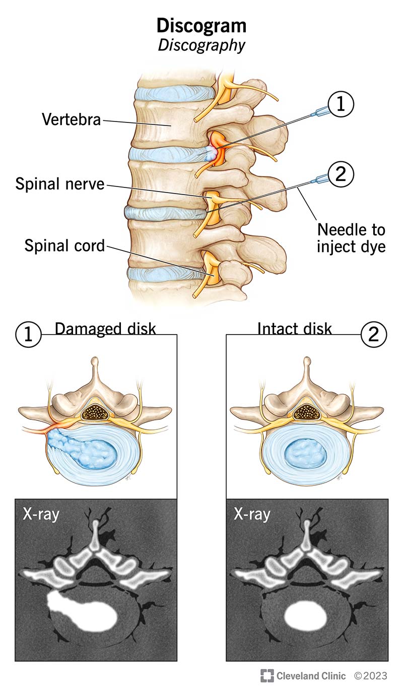
Image content: This image is available to view online.
View image online (https://my.clevelandclinic.org/-/scassets/images/org/health/articles/discogram)
A discogram (also called discography) is a type of imaging test that helps diagnose chronic (long-term) back pain. It may be able to tell your healthcare provider whether your symptoms result from a worn or damaged spinal disk.
Advertisement
Cleveland Clinic is a non-profit academic medical center. Advertising on our site helps support our mission. We do not endorse non-Cleveland Clinic products or services. Policy
You have disks between every vertebrae — the bones that make up your spine. The disks are like spongy cushions that absorb shock and keep your spinal bones from rubbing against each other. Damaged disks are a common cause of chronic (long-term) back pain.
Healthcare providers usually prefer other imaging tests — like CT (computed tomography) scans or MRI (magnetic resonance imaging) — to diagnose back pain. But they might need to take a discogram if:
Surgeons may also use discograms before spinal fusion surgery to confirm which disks need to be removed.
You might hear healthcare providers refer to cervical or lumbar discograms. These terms refer to the same procedure but specify which section of your spine needs imaging. If you have neck or upper back pain, you’ll need a cervical discogram. If you have lower back pain, you’ll need a lumbar discogram.
It’s important to note that damaged spinal disks don’t always cause back pain. For this reason, discograms are controversial in the medical community. But many healthcare providers believe that a discogram can help confirm suspected findings on a CT or MRI scan.
Advertisement
A discogram uses fluoroscopy to see how your vertebrae and spinal disks move. Fluoroscopy produces a moving X-ray image — a video you can watch in real time.
Your provider will also inject a contrast material (dye) into each disk to highlight potential areas of concern. If the contrast stays contained inside of your disk, it’s healthy. If the contrast spreads beyond the outer border of your disk, the disk might be worn or torn.
Your healthcare provider will give you instructions to follow before your discogram procedure. In general, you should:
When you arrive for your appointment, a provider will take you to an X-ray room where you’ll lie on an exam table. They’ll position you on your side and place pillows to keep you comfortable.
Most providers use IV (intravenous) sedation during a discogram. They’ll place an IV in a vein (usually in your arm or hand) and give you sedative medications to help you relax. They’ll also monitor your oxygen, blood pressure and heart rate throughout the procedure.
Once you’re comfortable, your provider will:
Your provider will use fluoroscopy to take images during the discogram procedure. Afterward, they may also take a CT scan or an MRI. Using more than one imaging test can help your provider make an accurate diagnosis.
From start to finish, a discogram usually takes about one hour.
The area near the injection sites might feel tender or uncomfortable for several hours after your discogram. You can manage this pain with at-home icing. Wrap an ice pack in a towel and place it over the sore areas for 20 minutes, then remove it for 20 minutes. Repeat as often as necessary. You can also take pain relievers like acetaminophen (Tylenol®) or ibuprofen (Motrin®).
Advertisement
Discogram complications are rare, but could include:
Most people can comfortably return to work, school and other routine activities after 24 hours. But it might take a few days for the soreness to go away.
A radiologist will read the results of your discogram to see if the contrast material stays inside each disk or if it leaks out.
If it stays inside your disk, that’s a normal (negative) result. If it leaks outside your disk, it indicates wear-and-tear damage (positive result).
Once your radiologist reviews your discogram, they’ll send the results to your provider. Once they have the results, your provider will discuss them with you. Most people get their results within a week.
It depends on your situation. Your provider may want to take additional imaging tests before deciding on a treatment plan. Or they may recommend spinal fusion — a procedure to remove the affected disk and fuse the vertebrae to reduce back pain symptoms.
After your discogram, call your healthcare provider if you develop:
Advertisement
If you have a damaged spinal disk, you’ll probably feel some pain when your provider injects the contrast material into it. Most people report the pain is very similar to the symptoms they experience every day. While temporarily unpleasant, this sensation can help your provider pinpoint which disks have wear and tear.
You may also have lingering discomfort for a few days. If this happens, take over-the-counter pain relievers and ice the area a few times each day.
Both are imaging tests providers use to help diagnose back pain, but they differ in a few ways:
| MRI | Discogram |
|---|---|
| Non-invasive. | Invasive. |
| No radiation. | Uses radiation. |
| May or may not use contrast material. | Uses contrast material. |
| Can detect issues with your vertebrae, soft tissues, ligaments, nerves and disks. | Can detect issues with your spinal disks. |
| MRI | |
| Non-invasive. | |
| Discogram | |
| Invasive. | |
| No radiation. | |
| Discogram | |
| Uses radiation. | |
| May or may not use contrast material. | |
| Discogram | |
| Uses contrast material. | |
| Can detect issues with your vertebrae, soft tissues, ligaments, nerves and disks. | |
| Discogram | |
| Can detect issues with your spinal disks. |
It’s common for healthcare providers to take CT scans or MRI scans first. If they need more information after trying other imaging tests, they may request a discogram.
Chronic back pain can seriously hinder your quality of life. And when you’ve tried several treatments with no improvement, it can feel frustrating, like you’ll never find a solution.
Not everyone with back pain needs a discogram. But in certain cases, this imaging test can help confirm worn spinal disks and rule out other conditions. If you’ve been living with back pain and nothing seems to help, ask your healthcare provider if a discogram could help with your diagnosis.
Advertisement


Sign up for our Health Essentials emails for expert guidance on nutrition, fitness, sleep, skin care and more.
Learn more about the Health Library and our editorial process.
Cleveland Clinic’s health articles are based on evidence-backed information and review by medical professionals to ensure accuracy, reliability and up-to-date clinical standards.
Cleveland Clinic’s health articles are based on evidence-backed information and review by medical professionals to ensure accuracy, reliability and up-to-date clinical standards.
When you need a clear picture of what’s happening inside your body, the Cleveland Clinic imaging team is here for you.
