The optic chiasm helps you see one clear image and perceive depth. This X-shaped structure is where the nerves from your eyes meet under your brain. But because of its location, it’s at risk of damage. This can cause changes in your vision.
Advertisement
Cleveland Clinic is a non-profit academic medical center. Advertising on our site helps support our mission. We do not endorse non-Cleveland Clinic products or services. Policy
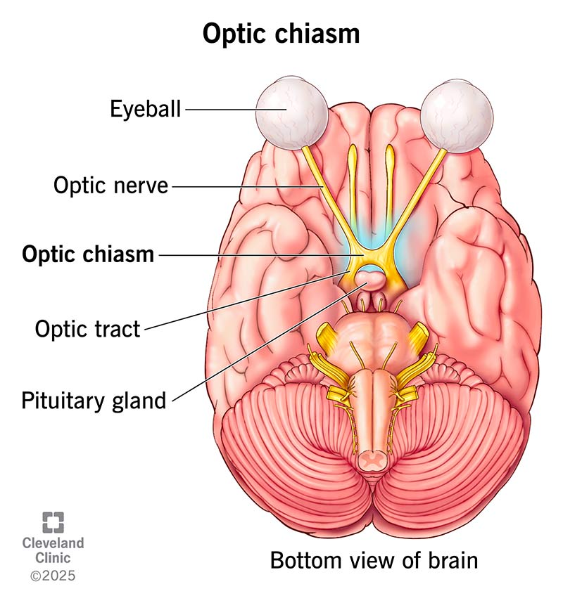
Image content: This image is available to view online.
View image online (https://my.clevelandclinic.org/-/scassets/images/org/health/articles/optic-chiasm)
The optic chiasm is where the optic nerves from each eye meet and cross. It’s responsible for transmitting visual information. This intersection helps your brain combine what both of your eyes see. Because of this meeting point, when you open your eyes, you get a single image.
Advertisement
Cleveland Clinic is a non-profit academic medical center. Advertising on our site helps support our mission. We do not endorse non-Cleveland Clinic products or services. Policy
The optic nerve is the cable that connects your eye to your brain. Both optic nerves meet and cross at the chiasm. It sits at the base of your brain. It’s surrounded by blood vessels and glands.
Because of its location, it’s at risk of damage to the surrounding glands or blood vessels. This might lead to vision changes or even vision loss.
The optic chiasm functions to:
Your optic chiasm is at the bottom of your brain. It sits below your hypothalamus and just above your pituitary gland. It’s in an area called the suprasellar cistern. This is close to your third ventricle (fluid-filled cavities). It’s also surrounded by a loop of arteries known as the circle of Willis.
Several important parts come together at the optic chiasm to help you see:
Advertisement
It has an “X” shape. “Chiasm” comes from the Greek letter “chi” (X). The shape happens when optic nerve fibers from both eyes cross. On average, it’s about a half inch or 13.5 millimeters wide.
Different conditions can press on the optic chiasm and affect your vision. These include:
Other conditions can affect how your optic chiasm works, including:
Damage may lead to vision changes, like:
Unless you’re at a busy intersection or knitting, you probably don’t think much about things crossing. But inside your brain, there’s one happening all the time. It’s how you see one world instead of two. As always, if anything about your vision seems off, let your provider know. They can check to see that your optic chiasm and its connected parts are working as they should.
Advertisement


Sign up for our Health Essentials emails for expert guidance on nutrition, fitness, sleep, skin care and more.
Learn more about the Health Library and our editorial process.
Cleveland Clinic’s health articles are based on evidence-backed information and review by medical professionals to ensure accuracy, reliability and up-to-date clinical standards.
Cleveland Clinic’s health articles are based on evidence-backed information and review by medical professionals to ensure accuracy, reliability and up-to-date clinical standards.
Getting an annual eye exam at Cleveland Clinic can help you catch vision problems early and keep your eyes healthy for years to come.
