A chest X-ray uses a focused beam of radiation to look at your heart, lungs and bones. Healthcare providers use chest X-rays to diagnose or treat conditions like pneumonia, emphysema or COPD. Chest X-rays are quick, noninvasive tests. Usually, you’ll know the results of your X-ray within one to two days.
Advertisement
Cleveland Clinic is a non-profit academic medical center. Advertising on our site helps support our mission. We do not endorse non-Cleveland Clinic products or services. Policy
A chest X-ray is a test that creates an image of your heart, lungs and bones. Other terms for chest X-ray are chest radiograph or CXR.
Advertisement
Cleveland Clinic is a non-profit academic medical center. Advertising on our site helps support our mission. We do not endorse non-Cleveland Clinic products or services. Policy
Chest X-rays help healthcare providers diagnose issues that cause symptoms in your heart or lungs. Some of these symptoms include:
Your healthcare provider may also recommend a chest X-ray to diagnose or monitor certain health conditions, including:
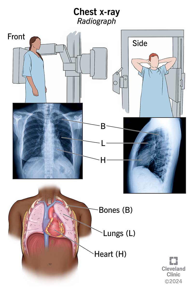
Image content: This image is available to view online.
View image online (https://my.clevelandclinic.org/-/scassets/images/org/health/articles/10228-chest-x-ray)
Your body’s tissues vary in thickness. When radiation passes through your body, each structure allows a different amount of radiation to pass through.
For example, your bones are very thick and don’t allow much radiation to pass through. Bones look white on an X-ray image. Your lungs, however, allow more radiation through. Your lungs look gray on an X-ray image.
Healthcare providers look at the colors and shading on an X-ray to diagnose and treat health conditions.
Chest X-rays require little to no preparation. When you get ready for your appointment, wear loose, comfortable clothing that doesn’t contain metal (zippers, snaps, bra closures) and leave jewelry at home.
If you have body piercings, ask your X-ray center for specific instructions. Body jewelry can interfere with clear images. You may need to remove them.
Advertisement
A radiology technologist will take your chest X-ray. These healthcare providers have specific training in X-ray testing.
You may change into a medical gown at your provider’s office. The X-ray technologist will also ask you to remove necklaces or any other metal around your chest area.
Typically, your chest X-ray consists of two parts:
During the chest X-ray, you need to remain very still and hold your breath. Any movement — even breathing in and out — can blur the X-ray image.
Chest X-rays usually take a few minutes to complete.
After the X-ray, your radiation technologist may ask you to wait a few minutes while they look at the images. If any of the images are blurry, they may have to retake the X-rays.
Next, the technologist will send your X-ray images to a radiologist who reviews them for normal and abnormal findings. Your healthcare provider will then review the images and radiologist’s report so they can discuss your X-ray results with you.
X-rays use a very small amount of radiation. Providers use only the necessary amount to capture quality images.
In nonemergency cases, you’ll probably get your X-ray results within one to two days. In an emergency, you’ll usually know your results in a few minutes or hours.
Your healthcare provider will review your chest X-ray results in detail and explain what they mean. In general:
If you get abnormal results, your provider may recommend additional tests (like a CT scan or PET scan) for more information.
Let your healthcare provider know if you have:
A chest X-ray usually consists of two images — one taken from the front and the other taken from the side. But in some cases, a technologist might take a total of four images, including one as you lie on your back and one as you lie on your side.
Advertisement
Always tell your healthcare provider if you might be pregnant — even if it’s only a small possibility. Radiation exposure can cause damage to a developing fetus.
The amount of radiation simple chest X-rays use is so small that it typically doesn’t cause issues during pregnancy. But healthcare providers prefer to limit radiation exposure until after your delivery. They’ll determine whether you need an X-ray based on your symptoms.
A chest X-ray is a quick, noninvasive way for your provider to check the overall health of your lungs, heart and ribcage. It’s often one of the first tests they use to diagnose broken bones or lung and heart conditions. And it can help your provider determine what kind of treatment you need.
Advertisement


Sign up for our Health Essentials emails for expert guidance on nutrition, fitness, sleep, skin care and more.
Learn more about the Health Library and our editorial process.
Cleveland Clinic’s health articles are based on evidence-backed information and review by medical professionals to ensure accuracy, reliability and up-to-date clinical standards.
Cleveland Clinic’s health articles are based on evidence-backed information and review by medical professionals to ensure accuracy, reliability and up-to-date clinical standards.
When you need a clear picture of what’s happening inside your body, the Cleveland Clinic imaging team is here for you.
