An arthrogram is a test that helps healthcare providers diagnose joint issues like hip or shoulder pain. These tests can show ligament, tendon and cartilage conditions in clear detail. An arthrogram involves injecting a contrast material directly into your joint before taking an X-ray, CT scan, MRI, ultrasound or another type of imaging test.
Advertisement
Cleveland Clinic is a non-profit academic medical center. Advertising on our site helps support our mission. We do not endorse non-Cleveland Clinic products or services. Policy
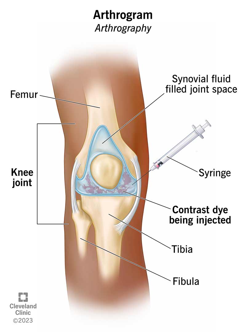
Image content: This image is available to view online.
View image online (https://my.clevelandclinic.org/-/scassets/images/org/health/articles/arthrogram)
An arthrogram is a medical imaging test. It uses contrast material (dye) to give healthcare providers a detailed view of what’s happening inside your joints.
Advertisement
Cleveland Clinic is a non-profit academic medical center. Advertising on our site helps support our mission. We do not endorse non-Cleveland Clinic products or services. Policy
Another name for arthrogram is arthrography.
Your provider may recommend an arthrogram if a physical exam or medical tests (like X-rays) don’t provide enough information for a diagnosis.
Providers also use arthrograms to:
There are several kinds of arthrograms based on the location of the affected area. Common types include:
Arthrogram is a two-part procedure. First, a trained healthcare provider injects a special dye (called contrast) into a vein or directly into your affected joint. The dye absorbs into your joint, making tiny structures (and hard-to-detect issues) easier to see.
Next, a provider takes pictures of your joint. To do this, your provider may use:
In some cases, your provider may take pictures of your joint before and after the dye injection.
Advertisement
There are a few things you can do to prepare for your arthrogram:
Your provider may ask you to remove clothing, depending on which joint they need to access.
A radiologist will perform your arthrogram. A specially trained technologist may assist in taking medical images. Your providers may take images (like X-rays) before and after the joint injection.
During an arthrogram, your provider will:
Providers often perform fluoroscopy and X-ray in the same room where you get the joint injection. If you need a CT or MRI, your provider may walk you to another room to take those images right after your joint injection.
It’s important to have pictures taken soon after the joint injection. Otherwise, the dye travels throughout your body. Once that happens, images no longer provide extra detail around your joint tissues.
During therapeutic arthrography, your provider injects medication (like cortisone) right inside your joint. The injection aims to reduce inflammation or relieve pain.
You should be able to resume normal activities shortly after the test is over. Some people have a bit of soreness or swelling around their joints after an arthrogram. If that’s the case, take it easy for the rest of the day.
Any discomfort should go away within two days. If it doesn’t, call your healthcare provider for guidance.
The overall risks are low. Possible arthrogram side effects include:
Advertisement
The radiologist who performed your arthrogram will review the pictures of your joint. They’ll send your results to your provider who ordered the study, usually within 24 hours after the test. Results may take longer if you have your test on a Friday.
Your provider will discuss your test results with you. They may recommend more tests or possible treatments (like joint replacement surgery) to fix the joint and relieve your symptoms.
You may feel slight discomfort when your provider releases the contrast material into the joint. Many people report feeling a “full” or “tight” sensation around the joint.
It depends on the type of imaging test your provider uses. Two of the most common contrast materials include iodine- and gadolinium-based dyes.
Generally, you can drive yourself home after your procedure. But if you plan to take sedative medications to help you relax, you’ll need to arrange for a trusted friend or family member to take you to and from your appointment. Ask your provider whether you’ll need sedation for your arthrogram.
Joint pain can keep you from living an active life. But diagnosing joint pain can be tricky. Arthrography reveals fine details of the tiny structures inside your joints. It’s a relatively painless diagnostic procedure with few risks. If pain in your hip, shoulder or another joint is holding you back, reach out to your provider about tests or treatments that may help you find relief.
Advertisement


Sign up for our Health Essentials emails for expert guidance on nutrition, fitness, sleep, skin care and more.
Learn more about the Health Library and our editorial process.
Cleveland Clinic’s health articles are based on evidence-backed information and review by medical professionals to ensure accuracy, reliability and up-to-date clinical standards.
Cleveland Clinic’s health articles are based on evidence-backed information and review by medical professionals to ensure accuracy, reliability and up-to-date clinical standards.
When you need a clear picture of what’s happening inside your body, the Cleveland Clinic imaging team is here for you.
