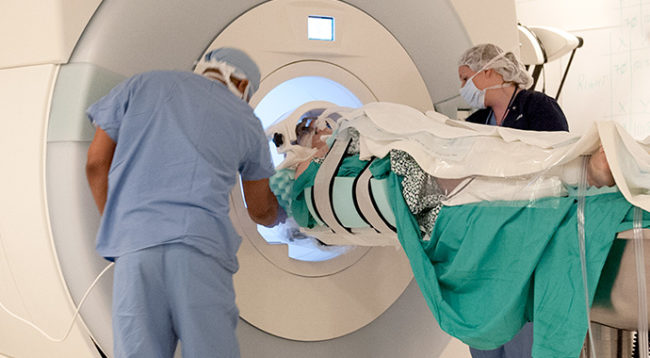
Patients with either essential tremor or Parkinson's disease associated tremor report over a 50 percent improvement in their tremors 3 months after the procedure.
Advertisement
Cleveland Clinic is a non-profit academic medical center. Advertising on our site helps support our mission. We do not endorse non-Cleveland Clinic products or services. Policy
MR-guided focused ultrasound is a treatment method that combines two technologies.
Advertisement
Cleveland Clinic is a non-profit academic medical center. Advertising on our site helps support our mission. We do not endorse non-Cleveland Clinic products or services. Policy
Magnetic resonance (MR) imaging aids vision and planning – the images provide surgeons with clear and highly detailed pictures, which helps pinpoint the area to treat and monitors treatment progress.
Ultrasound (sound waves) is a form of energy that can pass through various types of tissues – skin, fat, bone, and muscle. Highly focused ultrasound is the method of treatment. Guided by the MR images, over 1,000 beams of ultrasound are concentrated and focused on a specific target in the body. The beams raise the temperature of the targeted spot of tissue. The heat causes a burn that destroys the targeted tissue but does not damage the surrounding tissue.
The U.S. Food and Drug Administration has approved focused ultrasound for the following conditions:
Advertisement
Use of MR-guided focused ultrasound is being explored for other neurologic conditions including the tremors associated with multiple sclerosis, epileptic seizures that cannot be controlled by other treatment approaches, other movement disorders, stroke, brain tumors and neuropathic pain. Treating these other conditions is considered experimental at this time.
For essential tremor and Parkinson’s disease, over 1,000 highly focused beams of ultrasound are concentrated on a specific area in the brain’s thalamus. The thalamus in the brain is a relay station of motor and sensory signals. Essential tremor and Parkinson’s disease cause the thalamic circuitry to become abnormal, which results in tremors. The heat from the ultrasound causes a tiny burn or lesion on the targeted spot on the thalamus. Creating the tiny burn or lesion interrupts the abnormal activity, which relieves the tremors associated with these diseases.

Image content: This image is available to view online.
View image online (https://my.clevelandclinic.org/-/scassets/images/org/health/articles/21087-mr-guided-focused-ultrasound-for-treatment-of-tremor)
First, your head will be shaved on the morning of the procedure. This allows good contact between your head and the ultrasound. A urinary catheter may be placed to drain your bladder. Your vital signs (heart rate, blood pressure, oxygen level) will be monitored.
Your head will be placed in a head frame. This piece of equipment keeps your head steady and prevents it from moving. You will be given some medicine through an IV in your arm to keep you comfortable during this part of the procedure. Next, a silicone membrane will be placed on top of your head. The membrane seals the space where cold water will circulate between your scalp and the helmet of the ultrasound device. The water barrier helps keep your scalp cool and makes sure there is adequate contact between your head and the ultrasound equipment.
You will lie on an MRI bed, which slides in and out of the scanning area of the machine. The head frame is locked into position on the bed so you cannot move your head. Next, several scans will be taken of your brain. These scans help your physicians identify the area to be treated and the specific target area to aim the ultrasound waves.
Before treatment begins, you may be asked to draw spirals or write your name on a clipboard or perform certain hand and finger movements so your medical team can assess your tremor. You will be asked to repeat these tasks during various stages of treatment to see if the ultrasound is relieving your tremors.
Treatment begins with a number of short ultrasound pulses aimed at the target area. These low-energy (non-treatment level) pulses of ultrasound are administered to confirm that the proper target has been located. Once the accuracy of the target is confirmed by MR images, the ultrasound energy is gradually increased over a series of stages. At each stage, the temperature of the targeted tissue is checked and MR images confirm that the procedure is continuing as planned. The MR images provide real-time feedback so the surgeon can make any needed adjustments. You will be asked how you are feeling and will repeat hand and finger tasks to check treatment progress. Once your tremor is improving the ultrasound energy is increased until a small lesion is formed.
Advertisement
You will be awake during the entire procedure inside the MRI and will be able to speak to your medical team. You will be given an emergency stop button to hold during the procedure. If you are experiencing a problem or are concerned about how you are feeling, you can push the stop button at any time.
Once treatment is finished, more MR images will be taken and your urinary catheter, IV and all head gear will be removed.
The entire procedure, from preparation to getting off the table, takes about 3 to 4 hours.
You will move to a recovery room for observation. You may go home the same day or remain in the hospital for 24 to 48 hours. Your doctor will let you know when you can leave and when you need to return for a follow-up visit.
You should notice improvements while undergoing the procedure.
Essential tremor trial. In one of the pivotal safety and effectiveness trials leading to FDA approval, patients reported a 50 percent improvement in their tremors and motor functions 3 months after treatment compared to baseline and maintained a 40 percent improvement 1 year after treatment.
Tremor-dominant Parkinson’s disease. In the study that led to FDA approval, patients reported a 62 percent median improvement in their hand tremor 3 months after treatment compared with baseline.
Advertisement
Using this procedure:
The most common side effects include:
The side effects could start several days or weeks after treatment.
Risks and complications include:
Unfortunately no. The procedure does not treat the underlying disease nor prevent its progression.
Usually, you can return to normal activities within a few days.
It is best to speak with your doctors and to contact your insurance company directly to find out if this procedure is covered by your insurance.
Advertisement
This procedure may not be suitable if you:
Your medical team will conduct a complete assessment of your condition, conduct any needed addition tests, and then discuss if you are a candidate for this or other treatment options (such as deep brain stimulation).
Generally, MR-guided focused ultrasound may be an option if:

Sign up for our Health Essentials emails for expert guidance on nutrition, fitness, sleep, skin care and more.
Learn more about the Health Library and our editorial process.
Cleveland Clinic’s health articles are based on evidence-backed information and review by medical professionals to ensure accuracy, reliability and up-to-date clinical standards.
Cleveland Clinic’s health articles are based on evidence-backed information and review by medical professionals to ensure accuracy, reliability and up-to-date clinical standards.
Parkinson’s disease, essential tremor and dystonia are common movement disorders. And Cleveland Clinic has the expert care and support you need to manage them.
